. 2021 Sep;28(9):2971-2979.
doi: 10.1111/ene.14997. Epub 2021 Jul 16.

Bruns' nystagmus revisited: A sign of stroke in patients with the acute vestibular syndrome

Affiliations

Bruns' nystagmus revisited: A sign of stroke in patients with the acute vestibular syndrome

Georgios Mantokoudis et al. Eur J Neurol. 2021 Sep.

Abstract

Objective: Gaze-evoked nystagmus (GEN) is a central sign in patients with the acute vestibular syndrome (AVS); however, discriminating between a pathological and a physiologic GEN is a challenge. Here we evaluate GEN in patients with AVS.

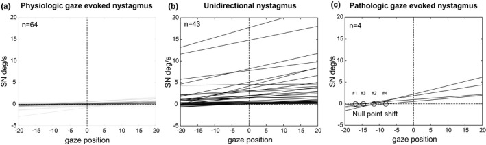
Methods: In this prospective cross-sectional study, we used video-oculography (VOG) to compare GEN in the light (target at 15° eccentric) in 64 healthy subjects with 47 patients seen in the emergency department (ED) who had AVS; 35 with vestibular neuritis and 12 with stroke. All patients with an initial non-diagnostic MRI received a confirmatory, delayed MRI as a reference standard in detecting stroke.

Results: Healthy subjects with GEN had a time constant of centripetal drift >18 s. VOG identified pathologic GEN (time constant ≤ 18 s) in 33% of patients with vestibular strokes, specificity was 100%, accuracy was 83%. Results were equivalent to examination by a clinical expert. As expected, since all patients with GEN had a SN in straight-ahead position, they showed the pattern of a Bruns' nystagmus.

Conclusions: One third of patients with AVS due to central vestibular strokes had a spontaneous SN in straight-ahead gaze and a pathological GEN, producing the pattern of a Bruns' nystagmus with a shift of the null position. The localization of the side of the lesion based on the null was not consistent, presumably because the circuits underlying gaze-holding are widespread in the brainstem and cerebellum. Nevertheless, automated quantification of GEN with VOG was specific, and accurately identified patients in the ED with AVS due to strokes.

Keywords: Bruns’ nystagmus; HINTS; acute vestibular syndrome; gaze-evoked nystagmus; gaze-holding nystagmus.


Conflict of interest statement

None of the investigators has any relevant financial interests, activities, relationships, or affiliations that represent a relevant financial conflict of interest with respect to the conduct or analysis of this study. TCS holds an endowed professorship sponsored by Touring Club Switzerland. The sponsor has no influence on the direction and content of the research conducted.

Figures

FIGURE 1
FIGURE 1
Box plots depicting the medians and the interquartile range (IQR) of physiologic GEN for left gaze (15 deg), straight ahead gaze (0 deg) and right gaze (−15 deg) in light in normal subjects. The circles and stars indicate outliers of 1.5*IQR and 3*IQR, respectively. Nystagmus slow‐phase velocity (SPV) is shown on the y‐axis, negative values indicate a right beating nystagmus
FIGURE 2
FIGURE 2
An example a pathologic GEN in a patient (#4) with a stroke involving the brainstem (right vestibular nucleus, posterior inferior medulla), both cerebellar tonsils and the inferior vermis (Figure 4, Pt. # 4). Panel (a) depicts horizontal eye positional data over time recorded with VOG. Note, left eye position is positive, right negative. The patient was first looking to the left with a left beating nystagmus (average 0.75 deg/s SPV), then he looked to the center, where nystagmus changed direction beating to the right. At right gaze (−10 deg), nystagmus intensity increased further but was still beating to the right. Panel (b) depicts the nystagmus intensity (slow‐phase velocity, deg/s) at all three gaze positions: right, center and left. The dotted line illustrates the regression line and its slope τau. The time constant was derived from the reciprocal of τau
FIGURE 3
FIGURE 3
The regression lines derived from gaze position plots from each subject. We reversed all regression lines into positive slopes in order to compare data. We separated data into three groups: Healthy subjects with physiologic GEN (Panel a), AVS patients with unidirectional nystagmus regardless of the underlying cause (Panel b) and AVS patients with pathologic GEN (Panel c). Note, that the regression line crossed the x‐axis defines the ‘null point’, at the gaze position with no nystagmus (‘null’ point). All four patients had an asymmetrical GEN with a shift of the null point. Note the data were mirrored for direction for comparison. Patients # 1 and 2 had shifts of the null to the right, Patient # 3 and 4# to the left
FIGURE 4
FIGURE 4
An MRI montage (axial MRI with diffusion‐weighted imaging) of stroke lesions in four patients (Pt) with pathologic GEN. We chose representative image sections for each patient. Lesions were located in the right midbrain for Pt #1, in the right midbrain and right superior cerebellar peduncle, tectum and right tegmentum for Pt #2, in the right cerebellar hemisphere (lateral) and right middle cerebellar peduncle for Pt #3, and in both cerebellar hemispheres, the vermis and the right posterior inferior medulla involving the right medial and lateral vestibular nuclei for Pt #4
FIGURE 5
FIGURE 5
Box plots depicting the medians and the interquartile range (IQR) of the time constant (Tc) recorded with VOG in healthy subjects (physiologic GEN) and in patients with pathologic GEN (Panel a). Outliers are not shown for illustrative purposes. Panel (b) shows a receiver operating characteristics curve (ROC) using the Tc as a predictive parameter for discrimination between pathologic GEN and physiologic GEN. A cut‐off value of <17.95 s favored a pathologic GEN (sensitivity 100%, specificity 84%)

References

    1. Leigh RJ, Zee DS. The Neurology of Eye Movements. Oxford, UK: Oxford University Press; 2015.
    1. Mantokoudis G, Wyss T, Zamaro E, et al. Stroke prediction based on the spontaneous nystagmus suppression test in Dizzy patients: a diagnostic accuracy study. Neurology. 2021;33986142. published online 2021. 10.1212/WNL.0000000000012176 - DOI - PMC - PubMed
    1. Kim HA, Yi HA, Lee H. Failure of fixation suppression of spontaneous nystagmus in cerebellar infarction: frequency, pattern, and a possible structure. Cerebellum. 2016;15:182‐189. - PubMed
    1. Kattah JC, Talkad AV, Wang DZ, Hsieh YH, Newman‐Toker DE. HINTS to diagnose stroke in the acute vestibular syndrome: three‐step bedside oculomotor examination more sensitive than early MRI diffusion‐weighted imaging. Stroke. 2009;40:3504‐3510. - PMC - PubMed
    1. Eggers SDZ, Bisdorff A, von Brevern M, et al. Classification of vestibular signs and examination techniques: Nystagmus and nystagmus‐like movements. J Vestib Res. 2019;29:57‐87. - PMC - PubMed

Publication types