

doi: 10.1161/CIRCRESAHA.121.319163.
Epub 2021 Jun 28.
Coronary Disease Association With ADAMTS7 Is Due to Protease Activity
Affiliations
- PMID: 34176299
- PMCID: PMC8349873
- DOI: 10.1161/CIRCRESAHA.121.319163
Item in Clipboard
Coronary Disease Association With ADAMTS7 Is Due to Protease Activity
Circ Res.
.
Abstract
[Figure: see text].
Keywords: atherosclerosis; coronary artery disease; phenotype; protease; wound healing.
Figures
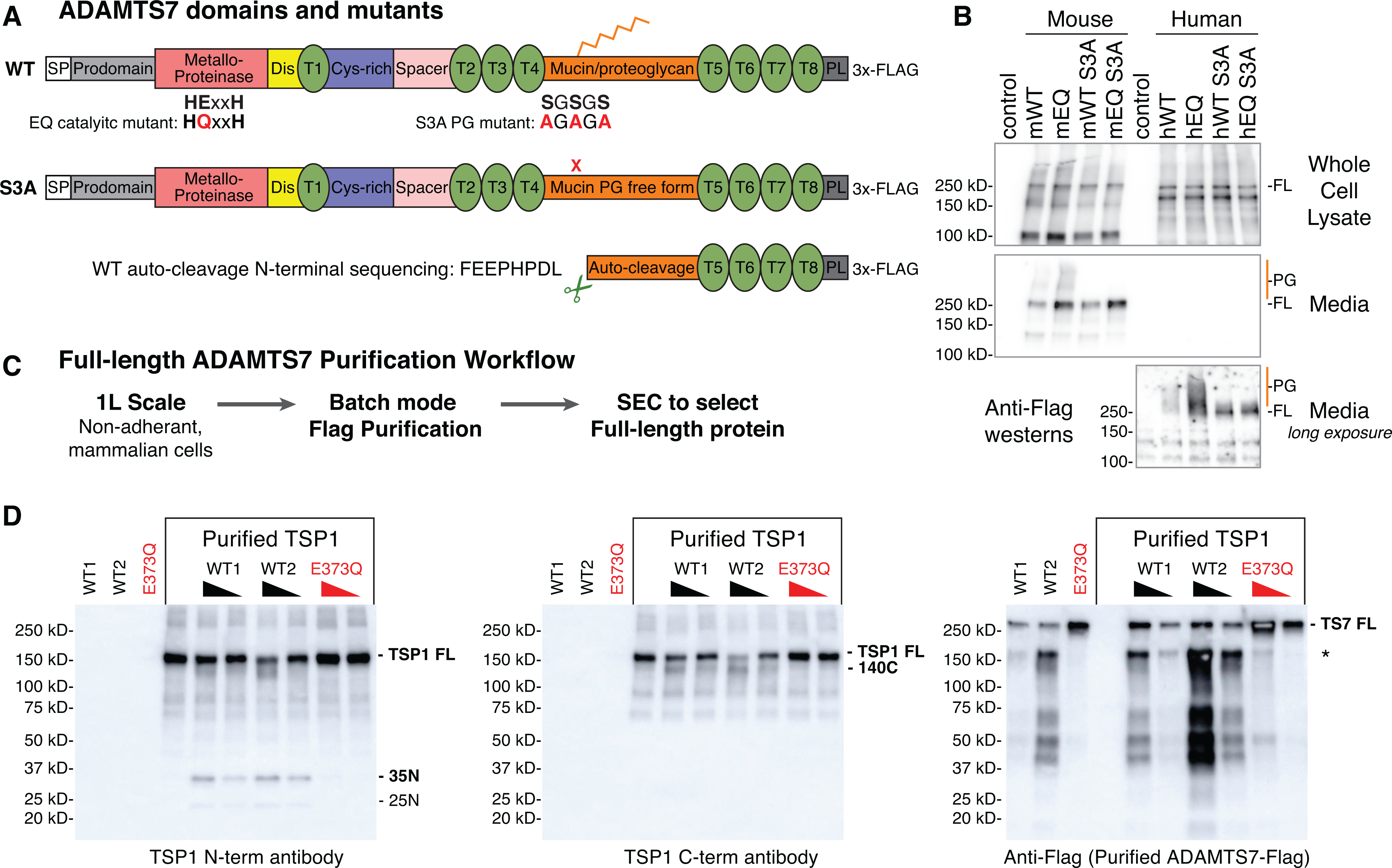
A, ADAMTS7 protein domains and locations of the glutamate to glutamine
(EQ) catalytic mutant and serine to alanine (S3A) substitutions to prevent
proteoglycan (PG) attachment. Abbreviated ADAMTS7 domains: signal peptide (SP),
disintegrin (Dis), thrombospondin repeats (T), cysteine-rich (Cys-rich),
protease and lacunin (PL). B, expression of full-length (FL) mouse and human
ADAMTS7 3xFlag proteins in the whole cell lysate and secreted in the media under
reducing conditions, detected by western blot using the M2-HRP antibody. High
molecular weight proteoglycan (PG) species are marked in orange. A longer
exposure of the conditioned media was required to visualize the secreted human
ADAMTS7 proteins. C, full-length ADAMTS7 two step purification workflow used to
purify WT S3A active enzyme and EQ S3A negative control protein. D,
Thrombospondin1 in vitro cleavage by purified full-length mouse
ADAMTS7 WT protein (* indicates co-purified auto-cleavage band). Western blots
under reducing conditions to resolve proteolytic TSP1 bands generated by active
ADAMTS7 WT purified enzyme. Presence of the E373Q catalytic mutation in the
purified full-length mouse ADAMTS7 EQ protein ablated the catalytic
activity.
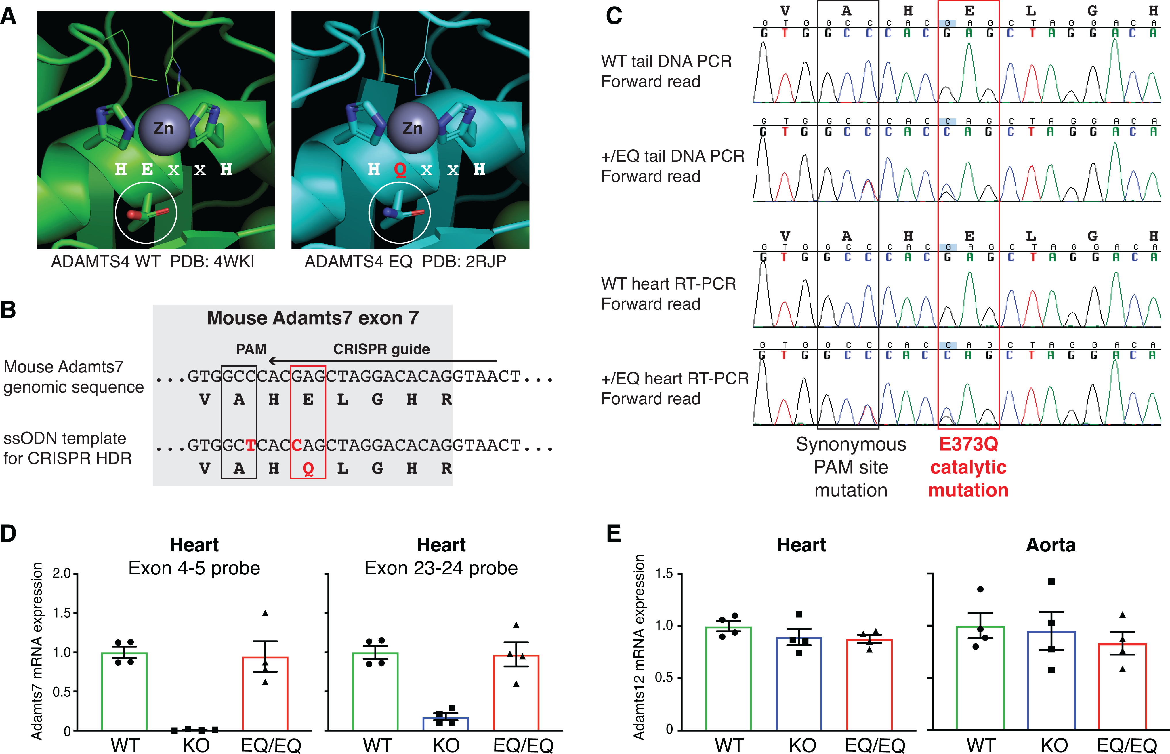
A, Structural context of the ADAMTS7 HExxH to HQxxH catalytic mutation.
Crystal structures from ADAMTS4 WT (PDB:4WKI) and ADAMTS4 EQ (PDB: 2RJP) were aligned and annotated in
PyMOL to highlight the catalytic residues and visualize the Zinc metal in the
active site. The E to Q substitution preserves the tertiary structure of ADAMTS4
and the residues “VAHELGH” in the active
site are conserved in ADAMTS7. B, Schematic of the Adamts7
E373Q catalytic mutant allele within exon 7. To generate the mutant allele, c.
1117G->C (p. E373Q) mutation at the Adamts7 catalytic
domain and c. 1113C->T (p. A371A) to disrupt the PAM site were induced by
CRISPR Homology-Directed Repair (HDR). C, Sanger sequencing of genomic tail DNA
PCR and heart mRNA RT-PCR from WT and heterozygous +/E373Q mice. Representative
forward reads show no evidence of allelic expression imbalance at the two
nucleotide substitutions. D, Adamts7 mRNA expression level in
the heart from WT, Adamts7 −/− (KO) and
homozygous E373Q/E373Q (EQ/EQ) mice were measured by real time quantitative
polymerase chain reaction (qPCR) using 2 TaqMan probe sets (exon 4–5
boundary and exon 23–24 boundary). n=4 per group. E,
Adamts12 mRNA expression levels were analyzed by real time
qPCR in the heart and aorta harvested from WT, KO and EQ/EQ mice. n=4 per group.
A Kruskal-Wallis test with Dunn’s multiple comparison test was applied to
D and E.
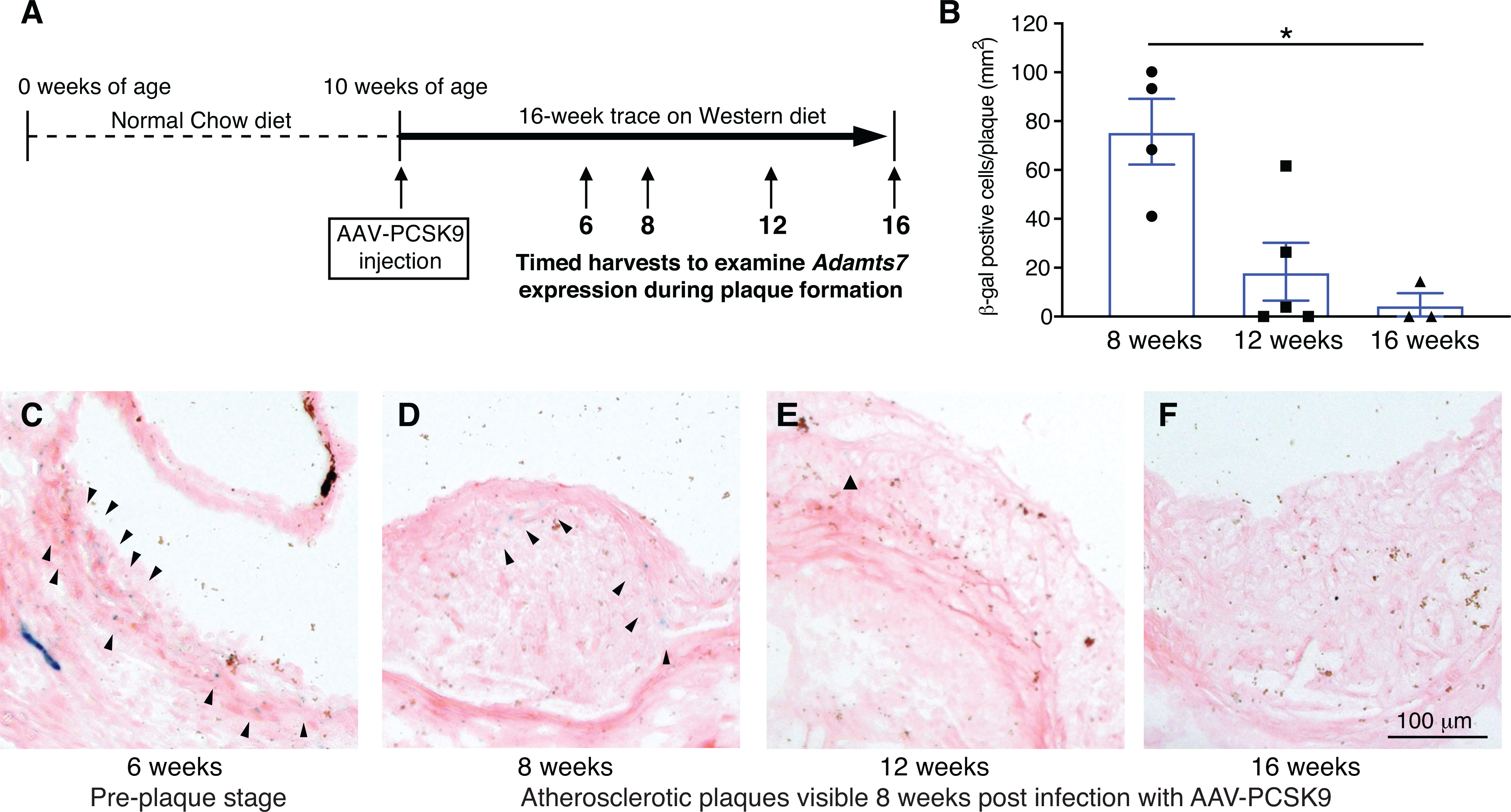
ADAMTS7 expression reported by the LacZ gene trap tm1b allele in
heterozygotes during atherogenesis at 6, 8, 12 and 16 weeks post infection with
AAV8-PCSK9. A, Stages examined in the atherogenic model post infection with
AAV8-PCSK9. B, Quantitative analysis of β-gal positive cells in the
plaque (n=3 to 5 per group, data points represent individual animals). A
Kruskal-Wallis test with Dunn’s multiple comparison test was applied,
*P<0.05. C-F, Representative photomicrographs of β-gal staining in
the aortic sinus at the indicated weeks after AAV8-PCSK9 injection.
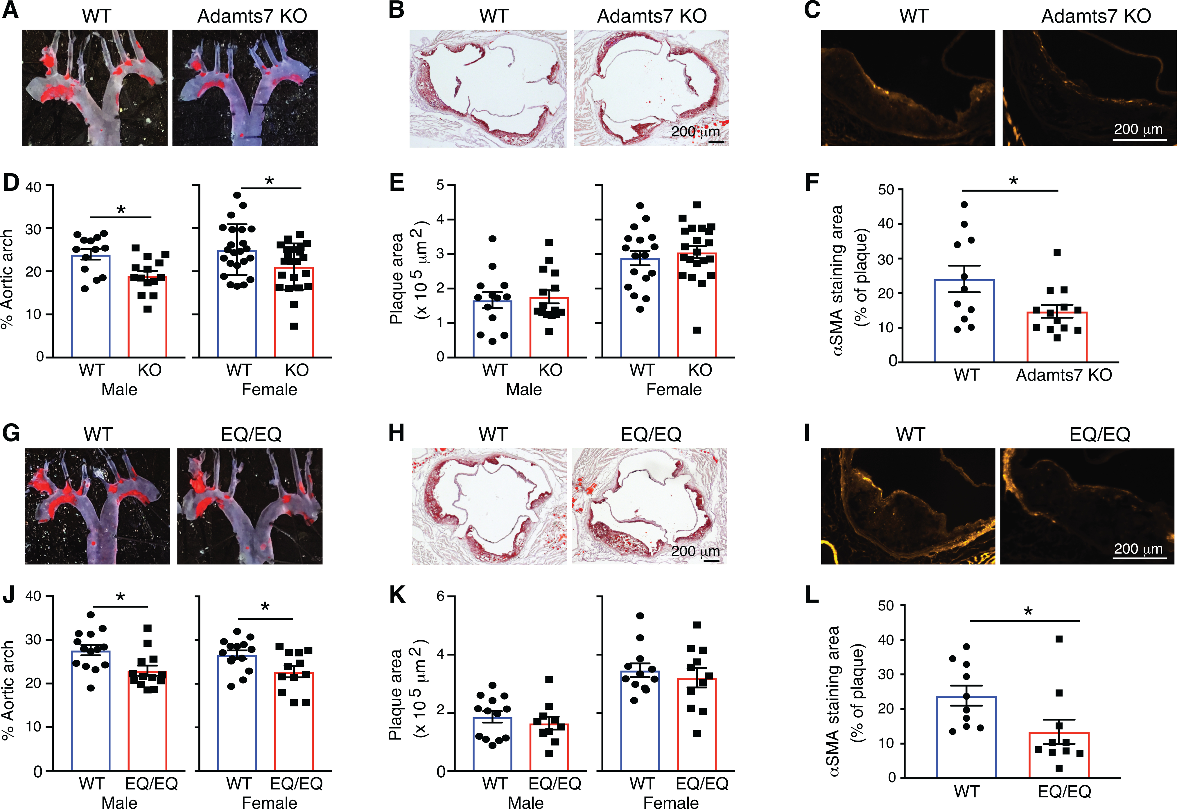
At the age of 10 weeks, mice were injected with rAAV8-D377Y-mPCSK9 and
challenged a western diet for an additional 16 weeks to stimulate
atherosclerosis. The mice were euthanized at 26 weeks of age for evaluation of
atherosclerosis in the aortic arch and aortic root. A, B, D and E,
Representative photomicrographs of Oil-Red O staining and quantitative analysis
of atherosclerotic lesion area in the aortas from littermate controls and
Adamts7 KO (D-E, males, n=13 to 15 per group; females, n=17
to 23 per group). G, H, J and K, From a separate +/EQ x +/EQ cross, littermate
controls and EQ/EQ homozygotes were evaluated for atherosclerosis (J-K, males,
n=10 to 14 per group; females, n=11 to 14 per group). C,F,I and L,
Representative sections and quantitative analyses of α-SMA positive
smooth muscle cells in the aortic sinus (n=10 to 12 per group). Data points
represent individual animals, error bars indicate means ±SEM. Two-way
ANOVA with Sidak’s multiple comparison test was applied to D, E, J and K.
A two-tailed Student’s test was applied to F, which showed normality and
Mann-Whitney test was applied to L, which did not pass a normality test.
Normality was tested by Kolmogorov-Smirnov test. *P<0.05.
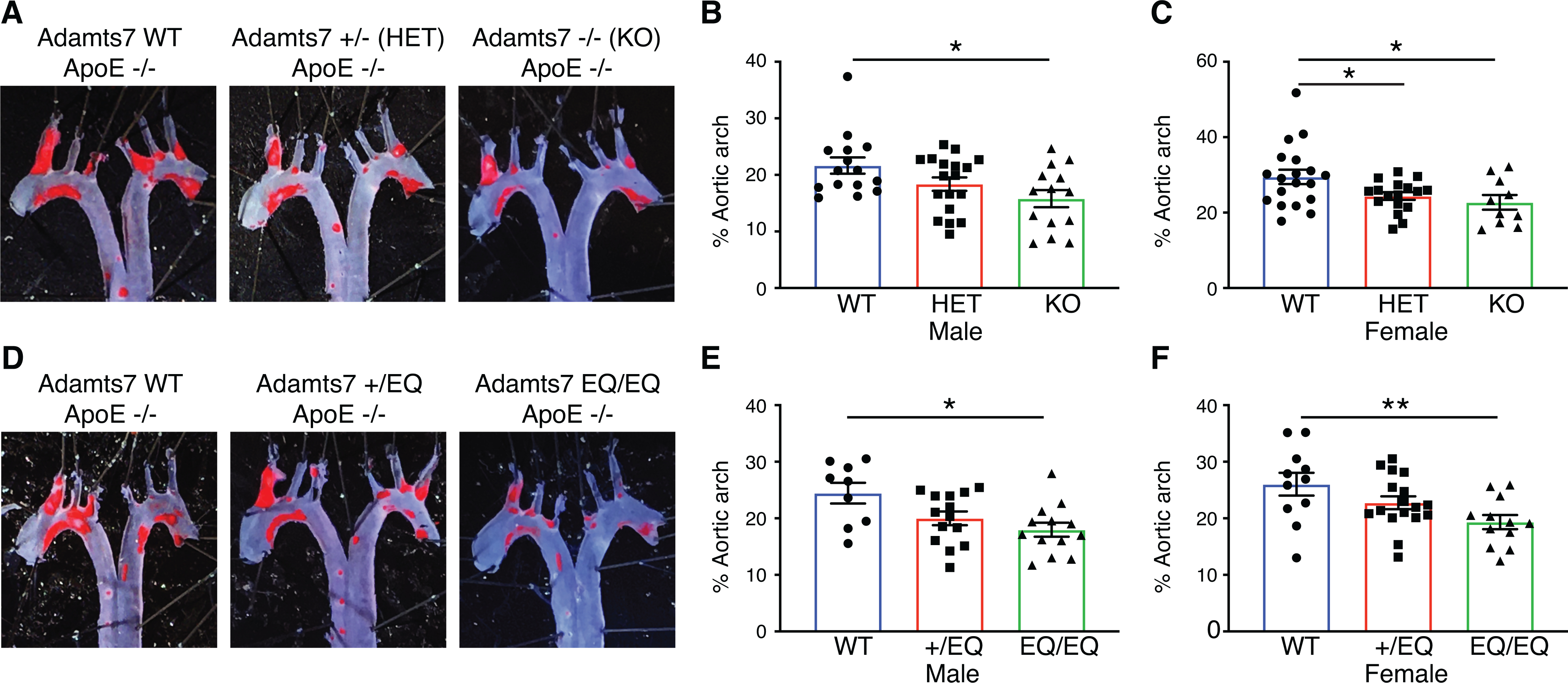
Mice from heterozygous intercrosses for the tm1b loss of function allele
or the E373Q catalytic mutant allele, were challenged with 10 weeks of high fat
diet to stimulate atherosclerosis on the ApoE KO background.
The mice were euthanized at 20 weeks of age for evaluation of atherosclerosis. A
and D, Representative photomicrographs of Oil-Red O staining and quantitative
analysis of atherosclerotic lesion area in the aortic arch. B, male WT vs
Adamts7 +/− (HET) vs Adamts7
−/− (KO), n=14 to 18 per group; C, female WT vs HET vs KO, n=10 to
19 per group; E, male WT vs +/EQ vs EQ/EQ, n=9 to 14 per group; F, female WT vs
+/EQ vs EQ/EQ, n=11 to 17 per each group. Data points represent individual
animals, error bars indicate means ±SEM, and two-way ANOVA with
Tukey’s test was applied. Normality was tested by Kolmogorov-Smirnov
test. *P<0.05, **P<0.01.
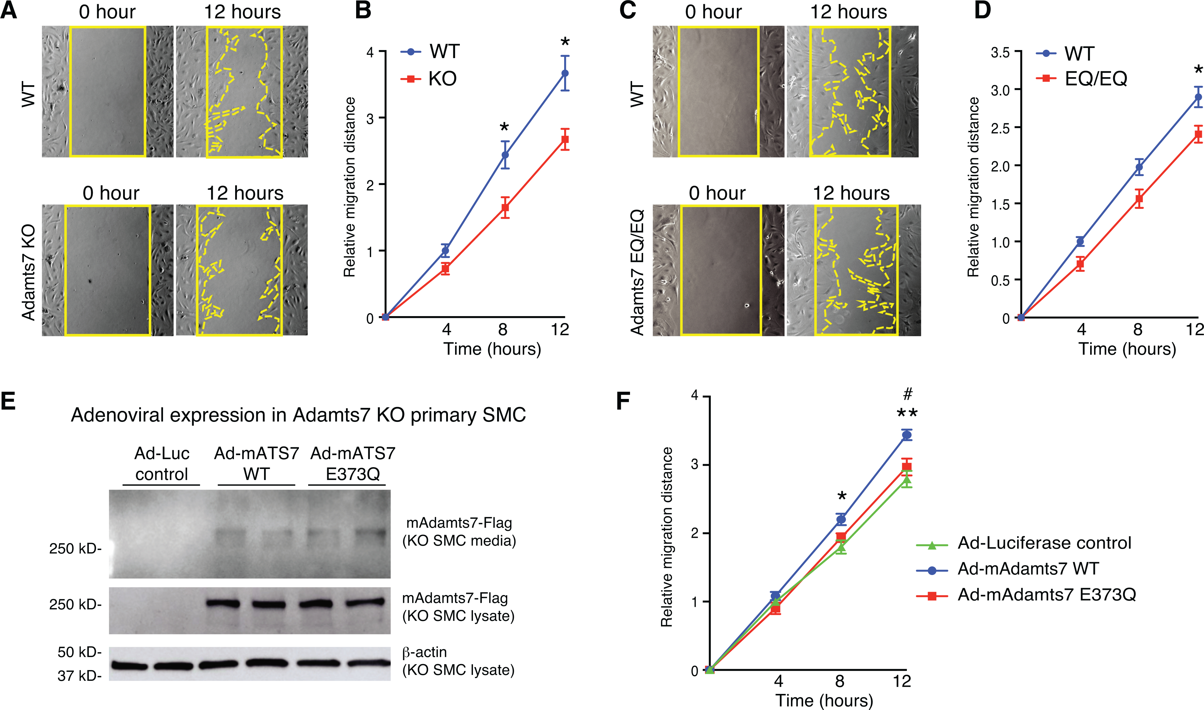
A-D, Migration of primary VSMCs from Adamts7
−/− (KO) and homozygous EQ/EQ catalytic mutant mice was assessed
by wound healing assay. A and C, Representative photomicrographs of the wound at
0 and 12 hours in indicated genotypes of VSMCs are shown. Solid line indicates
wound edge. Dotted line indicates migration edge. B and D, The distance of
migration is quantified at 4, 8 and 12 hours. n=8 to 9 per group. E,
Representative images of western blot in conditioned media and cell lysates from
adenovirus-infected primary Adamts7 KO VSMCs. Cell lysates and
conditioned media collected from Ad-Luciferase-, Ad-mAdamts7
(mAts7)-WT- and Ad-mAts7-E373Q-infected
primary Adamts7 KO VSMCs were subjected to western blot
analysis with anti-Flag antibody. F, Migration of primary
Adamts7 KO VSMCs infected with Ad-Luciferase,
Ad-mAdamts7 WT and Ad-E373Q was assessed by wound healing
assay. The mean distance of migration is quantified at 4, 8 and 12 hours. n= 8
to 9 per group. Error bars indicate ±SEM. Two-way RM ANOVA with
Sidak’s multiple comparisons test was applied to B, D. *P<0.05.
Two-way RM ANOVA with Tukey’s test was applied to F. Normality was tested
by Kolmogorov-Smirnov test. **P<0.01, (Ad-Luciferase vs Ad-mAts7 WT),
#P<0.05 (Ad-mAts7-WT vs Ad-mAts7–373Q).
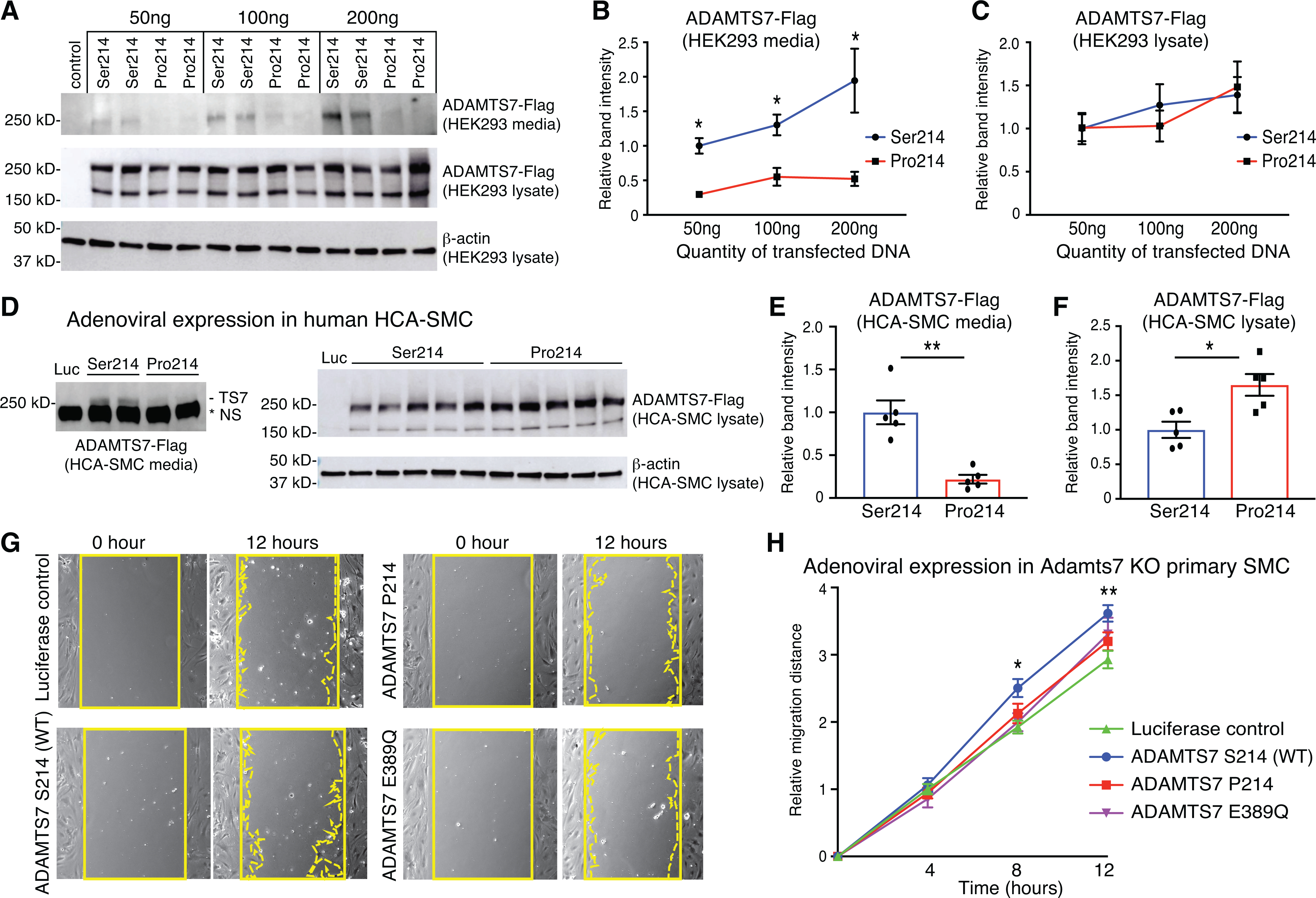
A-C, Representative images and quantifications of western blots from HEK
293 cells transfected with empty vector control, full-length ADAMTS7 risk
variant Ser214 or Pro214. A, Full-length ADAMTS7 3xFlag protein was detected by
anti-Flag antibodies from conditioned media and cell lysates. B-C, Relative band
intensities were quantified from n=4 per group. D-F, Representative images and
quantifications of western blot in conditioned media and cell lysates from
adenovirus-infected HCA-SMCs infected with Ad-Luciferase (Luc),
Ad-ADAMTS7-Ser214 or Ad-ADAMTS7-Pro214. D,
Full-length ADAMTS7 3xFlag protein was detected by western blot in conditioned
media and cell lysates. NS (non-specific) band migrating faster than
ADAMTS7-Flag is present in Luc control. E-F, Relative band intensities were
quantified (n=5 per group). Error bars indicate means ±SEM, Mann-Whitney
test was applied to B, C, E and F, *P<0.05, **P<0.01. G, Migration
of primary Adamts7 KO VSMCs infected with Ad-Luciferase,
Ad-ADAMTS7-Ser214 (WT), Ad-ADAMTS7-Pro214
or the Ad-ADAMTS7 catalytic mutant E389Q was assessed by wound
healing assay. Representative photomicrographs of the wound at 0 and 12 hours in
VSMCs infected with indicated adenovirus are shown. Solid line indicates wound
edge. Dotted line indicates migration edge. H, Migration was quantified at 4, 8
and 12 hours (n= 8 to 9 per group, two-way RM ANOVA with Tukey’s test.
Normality was tested by Kolmogorov-Smirnov test. *P<0.05, **P<0.01
for LUC vs Ser214).
Comment in
-
ADAMTS7 Knockdown in Context: Emerging Therapeutic Targets in Atherothrombosis.Circ Res. 2021 Aug 6;129(4):471-473. doi: 10.1161/CIRCRESAHA.121.319722. Epub 2021 Aug 5. Circ Res. 2021. PMID: 34351799 Free PMC article. No abstract available.
References
-
- Reilly MP, Li M, He J, Ferguson JF, Stylianou IM, Mehta NN, Burnett MS, Devaney JM, Knouff CW, Thompson JR, et al. Identification of ADAMTS7 as a novel locus for coronary atherosclerosis and association of ABO with myocardial infarction in the presence of coronary atherosclerosis: two genome-wide association studies. Lancet. 2011;377:383–92. - PMC - PubMed
-
- Coronary Artery Disease Genetics C. A genome-wide association study in Europeans and South Asians identifies five new loci for coronary artery disease. Nat Genet. 2011;43:339–44. - PubMed
Publication types
MeSH terms
Substances
Grants and funding
LinkOut - more resources
Full Text Sources
Other Literature Sources
Medical
Molecular Biology Databases
Research Materials
