. 2021 Apr-Jun;104(2):368504211026118.
doi: 10.1177/00368504211026118.

Protective effects of baicalin on caerulein-induced AR42J pancreatic acinar cells by attenuating oxidative stress through miR-136-5p downregulation

Affiliations

Protective effects of baicalin on caerulein-induced AR42J pancreatic acinar cells by attenuating oxidative stress through miR-136-5p downregulation

Zhu-Fen Zhao et al. Sci Prog. 2021 Apr-Jun.

Abstract

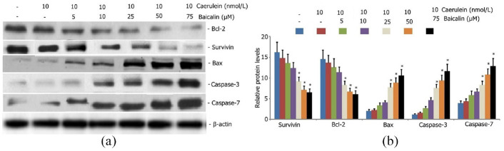
Baicalin, the main active component of Scutellaria baicalensis, has antioxidant and anti-apoptotic effects and is used to treat acute pancreatitis; however, its specific mechanism is unclear. This study aims to determine the protective effect and underlying mechanism of baicalin on AR42J pancreatic acinar cell injury. AR42J acinar cells (caerulein, 10 nmol/L) were induced in vitro to establish a cell model for acute pancreatitis. Cell relative survival was measured by thiazolyl blue tetrazolium bromide, and cell apoptosis and death were examined by flow cytometry. The expression levels of superoxide dismutase1 (SOD1), Bax, survivin, Bcl-2, caspase-3, and caspase-7 proteins were analyzed by Western blot, and those of SOD1 mRNA and miR-136-5p were determined by RT-PCR. The activities of GSH, SOD1, ROS, and MDA were also investigated. Compared with those of the caerulein group, the relative survival rate and activity of AR42J pancreatic acinar cells with different baicalin concentrations were significantly increased (p < 0.05), and the supernatant amylase level was markedly decreased (p < 0.05). In addition, the ROS and MDA activities and mir-136-5p expression were significantly decreased, and the GSH activities and SOD1 gene and protein expression levels were markedly increased (p < 0.05). These results suggest that baicalin reduced the caerulein-induced death of AR42J acinar cells and alleviated the caerulein-induced injury in pancreatic acinar cells by inhibiting oxidative stress. The mechanism may be related to the decreased expression of Mir-136-5p and the increased expression of SOD1 gene and protein.

Keywords: AR42J acinar cells; Baicalin; acute pancreatitis; miR-136-5p; oxidative stress; superoxide dismutase 1.


Conflict of interest statement

The author(s) declared no potential conflicts of interest with respect to the research, authorship, and/or publication of this article.

Figures

Figure 1.
Figure 1.
Effect of caerulein on AR42J pancreatic acinar cells viability, proliferation inhibition rate, apoptosis, and cells death rate. AR42J pancreatic acinar cells were incubated with various concentrations of caerulein (2.5, 5.0, 7.5, 10, and 15 nmol/L) for 24 h. AR42J pancreatic acinar cells viability was measured using CCK-8, proliferation inhibition rate was determined using MTT assay, and cells apoptosis and cells death rate was determined using flow cytometry. Data from three independent experiments are presented as mean ± SEM. *p < 0.05 versus the control group. #p < 0.05 versus the caerulein (7.5 nmol/L) group. p > 0.05 the caerulein (10 nmol/L) group versus the caerulein (15 nmol/L) group.
Figure 2.
Figure 2.
Effect of caerulein on oxidative stress in AR42J pancreatic acinar cells. AR42J pancreatic acinar cells were incubated with various concentrations of caerulein (2.5, 5.0, 7.5, 10, and 15 nmol/L) for 24 h. The activities of ROS, MDA, GSH, and SOD1 in AR42J pancreatic acinar cells were determined. Data from three independent experiments are presented as mean ± SEM. *p < 0.05 versus the control group. #p < 0.05 versus the caerulein (7.5 nmol/L) group. p > 0.05 the caerulein (10 nmol/L) group versus the caerulein (15 nmol/L) group.
Figure 3.
Figure 3.
Effect of caerulein on Bax, survivin, Bcl-2, caspase-3, caspase-7 protein expression in AR42J pancreatic acinar cells. AR42J pancreatic acinar cells were incubated with various concentrations of caerulein (2.5, 5.0, 7.5, 10, and 15 nmol/L) for 24 h. The activities of Bax, survivin, Bcl-2, caspase-3, caspase-7 protein expression in AR42J pancreatic acinar cells were determined by Western blot analysis. Data from three independent experiments are presented as mean ± SEM. *p < 0.05 versus the control group. #p < 0.05 versus the caerulein (7.5 nmol/L) group. p > 0.05 the caerulein (10 nmol/L) group versus the caerulein (15 nmol/L) group.
Figure 4.
Figure 4.
Effect of miR-136-5p inhibitor on AR42J pancreatic acinar cells apoptosis, decreased cells death rate, viability, and proliferation rate. The AR42J pancreatic acinar cells were transfected with miR-136-5p mimic and miR-136-5p inhibitor in the presence of 10 nmol/L caerulein for 24 h: (a and b) AR42J pancreatic acinar cells apoptosis and cells death rate was determined using flow cytometry, (c) AR42J pancreatic acinar cells viability was measured by CCK-8, and (d) AR42J pancreatic acinar cells proliferation rate was measured by MTT. The data from three independent experiments are presented as the mean ± SD. *p < 0.05 versus the miR-136-5p mimic control group. #p < 0.05 versus the miR-136-5p inhibitor control group.
Figure 5.
Figure 5.
Effect of miR-136-5p inhibitor on oxidative stress in AR42J pancreatic acinar cells. The AR42J pancreatic acinar cells were transfected with miR-136-5p mimic and miR-136-5p inhibitor in the presence of 10 nmol/L caerulein for 24 h. The activities of ROS, MDA, GSH, and SOD1 in AR42J pancreatic acinar cells were determined. Data from three independent experiments are presented as mean ± SEM. AR42J pancreatic acinar cells proliferation rate was measured by MTT. The data from three independent experiments are presented as the mean ± SD. *p < 0.05 versus the miR-136-5p mimic control group. #p < 0.05 versus the miR-136-5p inhibitor control group.
Figure 6.
Figure 6.
Effect of miR-136-5p inhibitor on miR-136-5p and SOD1 expression in AR42J pancreatic acinar cells. The AR42J pancreatic acinar cells were transfected with miR-136-5p mimic and miR-136-5p inhibitor in the presence of 10 nmol/L caerulein for 24 h. The levels of miR-136-5p expression in AR42J pancreatic acinar cells were determined by RT-PCR. The levels of SOD expression in AR42J pancreatic acinar cells were determined by Western blot. Data from three independent experiments are presented as mean ± SD. *p < 0.05 versus the control group. #p < 0.05 versus the model group. *p < 0.05 versus the miR-136-5p mimic control group. #p < 0.05 versus the miR-136-5p inhibitor control group.
Figure 7.
Figure 7.
Effect of baicalin on AR42J pancreatic acinar cells oxidative stress, viability, apoptosis, and death rate. AR42J pancreatic acinar cells were incubated with various concentrations of baicalin (10, 25, 50, and 75 μM) for 24 h. AR42J pancreatic acinar cells apoptosis and cells death rate was determined using flow cytometry. AR42J pancreatic acinar cells viability was measured by CCK-8. The activities of ROS, MDA, and SOD1 in AR42J pancreatic acinar cells were determined. Data from three independent experiments are presented as mean ± SD. p > 0.05 versus the control group.
Figure 8.
Figure 8.
Effect of baicalin on the mRNA expression of miR-136-5p and SOD1 and the protein expression Bax, Bcl-2, caspase-3, and caspase-7 protein in AR42J pancreatic acinar cells. AR42J pancreatic acinar cells were incubated with various concentrations of baicalin (10, 25, 50, and 75 μM) for 24 h. The mRNA expression of miR-136-5p and SOD1 in AR42J pancreatic acinar cells was measured by RT-PCR, the protein expression Bax, Bcl-2, caspase-3, and caspase-7 protein in AR42J pancreatic acinar cells were measured by Western blotting. Data from three independent experiments are presented as mean ± SD. p > 0.05 versus the control group.
Figure 9.
Figure 9.
Effect of baicalein on miR-136-5p and SOD1 mRNA and protein expression in caerulein induced AR42J pancreatic acinar cells: (a) effect of caerulein on miR-136-5p and SOD1 mRNA and protein expression in caerulein induced AR42J pancreatic acinar cells. AR42J pancreatic acinar cells were incubated with various concentrations of caerulein (2.5, 5.0, 7.5, 10, and 15 nmol/L) for 24 h. miR-136-5p and SOD1 mRNA expression were measured by RT-PCR. SOD1 protein expression by Western blot analysis. Data from three independent experiments are presented as mean ± SEM. *p < 0.05 versus the control group. #p < 0.05 versus the caerulein (7.5 nmol/L) group. p > 0.05 the caerulein (10 nmol/L) group versus the caerulein (15 nmol/L) group. (b) Effect of Baicalein on miR-136-5p and SOD1 mRNA and protein expression in caerulein induced AR42J pancreatic acinar cells. AR42J pancreatic acinar cells were incubated with caerulein (10 nmol/L) and various concentrations of baicalin (5, 10, 25, 50, and 75 μM) for 24 h. miR-136-5p and SOD1 mRNA expression were measured by RT-PCR. SOD1 protein expression by Western blot analysis. Data from three independent experiments are presented as mean ± SEM. (a) *p < 0.05; **p < 0.01 versus the control group. (b) *p < 0.05; **p < 0.01 versus the control group; #p < 0.05; ##p < 0.01versus the only caerulein group.
Figure 10.
Figure 10.
Effect of baicalein on oxidative stress in caerulein induced AR42J pancreatic acinar cells. AR42J pancreatic acinar cells were incubated with caerulein (10 nmol/L) and various concentrations of baicalin (5, 10, 25, 50, and 75 μM) for 24 h. ROS, MDA, GSH, and SOD1 in AR42J pancreatic acinar cells apoptosis was determined. Data from three independent experiments are presented as mean ± SD. *p < 0.05 versus the control group. #p < 0.05 versus the baicalin (25 μM) group. p > 0.05 the baicalin (50 μM) group versus the baicalin (75 μM) group.
Figure 11.
Figure 11.
Effect of baicalein on survivin, Bax, Bcl-2, caspase-3, caspase-7 protein expression in caerulein induced AR42J pancreatic acinar cells. AR42J pancreatic acinar cells were incubated with caerulein (10 nmol/L) and various concentrations of baicalin (5, 10, 25, 50, and 75 μM) for 24 h. Survivin, Bax, Bcl-2, caspase-3, caspase-7 protein expression levels were measured by Western blot analysis. Data from three independent experiments are presented as mean ± SEM. *p < 0.05. **p < 0.01 versus the only caerulein group.
Figure 12.
Figure 12.
Effect of baicalin on AR42J pancreatic acinar cells supernatant amylase, viability, proliferation inhibition rate, apoptosis, and death rate. AR42J pancreatic acinar cells were incubated with caerulein (10 nmol/L) and various concentrations of baicalin (5, 10, 25, 50, and 75 μM) for 24 h: (a) culture medium supernatant amylase AMS was measured, (b) AR42J pancreatic acinar cells viability was measured using CCK-8, (c) AR42J pancreatic acinar cells proliferation inhibition rate was determined using MTT, (d and e) AR42J pancreatic acinar cells apoptosis and death rate was determined using flow cytometry. Data from three independent experiments are presented as mean ± SEM. *p < 0.05; **p < 0.01 versus the control group. #p < 0.05; ##p < 0.01 versus the only caerulein group.
Figure 13.
Figure 13.
SOD1 is a downstream target of miR-136-5p: (a) decreased mRNA levels of SOD1 in caerulein-induced AR42J pancreatic acinar cells after miR-136-5p overexpression (p < 0.05). Levels of SOD1 mRNA were enhanced after miR-136-5p downregulation (p < 0.05), (b and c) levels of SOD1 protein was decreased in caerulein-induced AR42J pancreatic acinar cells with miR-136-5p overexpression relative to the empty vector control (p < 0.05). Levels of SOD1 protein was significantly increased in cells with miR-136-5p downregulation. (d) miR-136-5p was bound to the 3′-UTR regions of SOD1. Binding was interrupted in mutant SOD1. (e) Dual luciferase reporter assay indicated that miR-136-5p mimic was bound to the 3′-UTR region of the wild-type SOD1, rather than to SOD1 mutants (p < 0.05).

Similar articles

Cited by

References

    1. Amas Gómez L, Zubia Olaskoaga F. Results of the modification of an acute pancreatitis management protocol in Intensive Care medicine. Med Intensiva 2019; 43(9): 546–555. - PubMed
    1. Hines OJ, Pandol SJ. Management of severe acute pancreatitis. BMJ 2019; 367: l6227. - PubMed
    1. Ma Z, Song G, Zhao D, et al.. Bone marrow-derived mesenchymal stromal cells ameliorate severe acute pancreatitis in rats via hemeoxygenase-1-mediated anti-oxidant and anti-inflammatory effects. Cytotherapy 2019; 21(2): 162–174. - PubMed
    1. Lv C, Jin Q. Maresin-1 inhibits oxidative stress and inflammation and promotes apoptosis in a mouse model of caerulein-induced acute pancreatitis. Med Sci Monit 2019; 25: 8181–8189. - PMC - PubMed
    1. Máté G, Gazdag Z, Mike N, et al.. Regulation of oxidative stress-induced cytotoxic processes of citrinin in the fission yeast Schizosaccharomyces pombe. Toxicon 2014; 90: 155–166. - PubMed

Publication types

MeSH terms