

Primary CNS Extranodal Marginal Zone B-Cell Lymphoma: A Case Series of 2 Patients Treated with External Beam Radiation Therapy
- PMID: 34177522
- PMCID: PMC8216028
- DOI: 10.1159/000515780
Primary CNS Extranodal Marginal Zone B-Cell Lymphoma: A Case Series of 2 Patients Treated with External Beam Radiation Therapy
Abstract
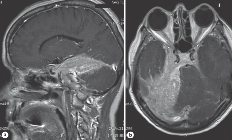
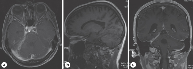
Primary CNS extranodal marginal zone B-cell lymphoma (MZBL) is a rare low-grade non-Hodgkin lymphoma characterized predominantly by small B cells, plasma cells, monocytoid cells, and scattered large immunoblasts. As a slow-growing tumor that remains localized, primary CNS MZBL carries an excellent clinical prognosis. Here, we report two cases of primary CNS MZBL successfully treated using external beam radiation therapy along with a literature review.
Keywords: B cell; Central nervous system lymphoma; Extranodal marginal zone lymphoma; Marginal zone B-cell lymphomas; Radiation therapy.
Copyright © 2021 by S. Karger AG, Basel.
Conflict of interest statement
The authors have no conflicts of interest to declare.
Figures


Similar articles
-
Unusual localization and clinical presentation of primary central nervous system extranodal marginal zone B‑cell lymphoma: A case report.Oncol Lett. 2023 Aug 4;26(3):408. doi: 10.3892/ol.2023.13994. eCollection 2023 Sep. Oncol Lett. 2023. PMID: 37600340 Free PMC article.
-
Exclusive detection of the t(11;18)(q21;q21) in extranodal marginal zone B cell lymphomas (MZBL) of MALT type in contrast to other MZBL and extranodal large B cell lymphomas.Am J Pathol. 1999 Dec;155(6):1817-21. doi: 10.1016/S0002-9440(10)65499-5. Am J Pathol. 1999. PMID: 10595910 Free PMC article.
-
Extranodal marginal zone B-cell lymphoma of the skin: a morphologic and immunophenotypic study of 11 cases.Am J Dermatopathol. 2000 Jun;22(3):205-11. doi: 10.1097/00000372-200006000-00001. Am J Dermatopathol. 2000. PMID: 10871062
-
Extranodal Marginal Zone B-cell Lymphoma of the Ocular Adnexa.Cancer Control. 2016 Apr;23(2):140-9. doi: 10.1177/107327481602300208. Cancer Control. 2016. PMID: 27218791 Review.
-
Extranodal Marginal Zone Lymphoma of the Central Nervous System.Clin Lymphoma Myeloma Leuk. 2018 Jan;18(1):34-37.e8. doi: 10.1016/j.clml.2017.09.012. Epub 2017 Sep 23. Clin Lymphoma Myeloma Leuk. 2018. PMID: 29103980 Review.
Cited by
-
Diagnosis and Treatment of Low-Grade Marginal Zone B-cell Lymphoma With Psychiatric Overlap.Cureus. 2024 May 6;16(5):e59735. doi: 10.7759/cureus.59735. eCollection 2024 May. Cureus. 2024. PMID: 38841029 Free PMC article.
-
Primary cerebral immunoglobulin light chain amyloidoma in a patient with multiple sclerosis.BMJ Case Rep. 2024 Jan 24;17(1):e256537. doi: 10.1136/bcr-2023-256537. BMJ Case Rep. 2024. PMID: 38272520
-
Primary extranodal marginal zone mucosa-associated lymphoid tissue-type B-cell lymphoma involving the dura: A case report.Surg Neurol Int. 2024 Mar 29;15:113. doi: 10.25259/SNI_792_2023. eCollection 2024. Surg Neurol Int. 2024. PMID: 38628522 Free PMC article.
-
Unusual localization and clinical presentation of primary central nervous system extranodal marginal zone B‑cell lymphoma: A case report.Oncol Lett. 2023 Aug 4;26(3):408. doi: 10.3892/ol.2023.13994. eCollection 2023 Sep. Oncol Lett. 2023. PMID: 37600340 Free PMC article.
References
-
- Zucca E, Arcaini L, Buske C, Johnson PW, Ponzoni M, Raderer M, et al. Marginal zone lymphomas: ESMO clinical practice guidelines for diagnosis, treatment and follow-up. Ann Oncol. 2020 01;31((1)):17–29. - PubMed
-
- Goda JS, Gospodarowicz M, Pintilie M, Wells W, Hodgson DC, Sun A, et al. Long-term outcome in localized extranodal mucosa-associated lymphoid tissue lymphomas treated with radiotherapy. Cancer. 2010 Aug;116((16)):3815–24. - PubMed
-
- Pavlou G, Pal D, Bucur S, Chakrabarty A, van Hille PT. Intracranial non-Hodgkin's MALT lymphoma mimicking a large convexity meningioma. Acta Neurochir. 2006 Jul;148((7)):791–3. discussion 93. - PubMed
Publication types
LinkOut - more resources
Full Text Sources
Research Materials
