

Myopic Simple Hemorrhage Presenting as Radial Hemorrhage in Henle's Fiber Layer
- PMID: 34177540
- PMCID: PMC8216004
- DOI: 10.1159/000510626
Myopic Simple Hemorrhage Presenting as Radial Hemorrhage in Henle's Fiber Layer
Abstract
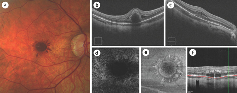
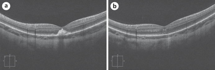
This case report describes a simple hemorrhage (SH) presenting as radial hemorrhage in Henle's fiber layer (HFL) in a patient with high myopia. A 26-year-old girl with high myopia was referred to our center for sudden onset of decreased vision and a central scotoma in the right eye (OD). Best corrected visual acuity (BCVA) was 20/100 OD. Fundus examination showed a stellate intraretinal hemorrhage in the fovea of the OD. The hemorrhage was organized in a peculiar petaloid pattern with feathery distal edges, suggesting localization within the radially oriented HFL. The presence of both choroidal neovascularization and microvascular abnormalities consistent with macular telangiectasia type 2 (MacTel 2) were excluded. Based on these findings, a diagnosis of myopic SH was made. At 4-month follow-up BCVA OD spontaneously improved to 20/40, without any treatment been ever administered to the patient. Spectral-domain optical coherence tomography OD showed reabsorption of the hemorrhage and almost complete restoration of the foveal architecture. The intraretinal location and spread of the hemorrhage into the HFL in our patient are an unusual presentation of SH, which vividly highlights the anatomy of the fovea. Since fibers in HFL are quite delicate and loosely arranged, this layer is very susceptible to deposition of transudates, exudates, hemorrhage, and other products. Radial hemorrhage in HFL has been originally reported in 4 patients as complication of MacTel 2. It has been previously postulated that it may represent a characteristic finding in MacTel 2 that may develop as a result of microvascular abnormalities of the deep retinal capillary plexus. On the contrary, our data suggest that radial hemorrhage in the HFL does not represent a characteristic finding of MacTel 2, but must rather be considered a non-specific sign with multiple possible etiologies.
Keywords: Eye disease; Henle fiber layer hemorrhage; Imaging; Macula/retina/vitreous; Maculopathy; Myopia; Myopic hemorrhage; Ophthalmoscopy; Radial hemorrhage; Retina; Simple hemorrhage.
Copyright © 2021 by S. Karger AG, Basel.
Conflict of interest statement
The authors have no conflicts of interest to declare.
Figures
Similar articles
-
Radial Hemorrhage in Henle Layer in Macular Telangiectasia Type 2.JAMA Ophthalmol. 2018 Oct 1;136(10):1182-1185. doi: 10.1001/jamaophthalmol.2018.2979. JAMA Ophthalmol. 2018. PMID: 30054619 Free PMC article.
-
Henle fibre layer haemorrhage: clinical features and pathogenesis.Br J Ophthalmol. 2021 Mar;105(3):374-380. doi: 10.1136/bjophthalmol-2019-315443. Epub 2020 May 6. Br J Ophthalmol. 2021. PMID: 32376610
-
Outer retinal deformity detected by optical coherence tomography in eyes with foveal hypoplasia.Graefes Arch Clin Exp Ophthalmol. 2016 Nov;254(11):2197-2201. doi: 10.1007/s00417-016-3385-z. Epub 2016 May 23. Graefes Arch Clin Exp Ophthalmol. 2016. PMID: 27216762
-
The OCT angular sign of Henle fiber layer (HFL) hyperreflectivity (ASHH) and the pathoanatomy of the HFL in macular disease.Prog Retin Eye Res. 2023 Jul;95:101135. doi: 10.1016/j.preteyeres.2022.101135. Epub 2022 Nov 1. Prog Retin Eye Res. 2023. PMID: 36333227 Review.
-
MACULAR TELANGIECTASIA TYPE 2 IN AN OTHERWISE HEALTHY TEENAGE BOY WITH CONSANGUINEOUS PARENTS.Retin Cases Brief Rep. 2018 Summer;12(3):200-203. doi: 10.1097/ICB.0000000000000481. Retin Cases Brief Rep. 2018. PMID: 27828905 Review.
References
-
- Rahimy E, Vander J. I see a seashell in my right eye. JAMA Ophthalmol. 2014 Dec;132((12)):1413. - PubMed
-
- Asai T, Ikuno Y, Nishida K. Macular microstructures and prognostic factors in myopic subretinal hemorrhages. Invest Ophthalmol Vis Sci. 2014 Jan 9;55((1)):226–32. - PubMed
-
- Nauman GOH, Apple DJ. Pathology of the eye. New York, NY: Springer-Verlag; 1985. Microscopic anatomy of the eye; p. p. 43.
-
- Baumal CR, Sarraf D, Bryant T, Gui W, Muakkassa N, Pichi F, et al. Henle fibre layer haemorrhage: clinical features and pathogenesis. Br J Ophthalmol. 2021;105((3)):374–80. - PubMed
Publication types
LinkOut - more resources
Full Text Sources
