

Management of Retinal Detachment Associated with Morning Glory Disc Syndrome
- PMID: 34177542
- PMCID: PMC8215972
- DOI: 10.1159/000516205
Management of Retinal Detachment Associated with Morning Glory Disc Syndrome
Abstract
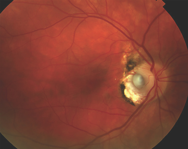
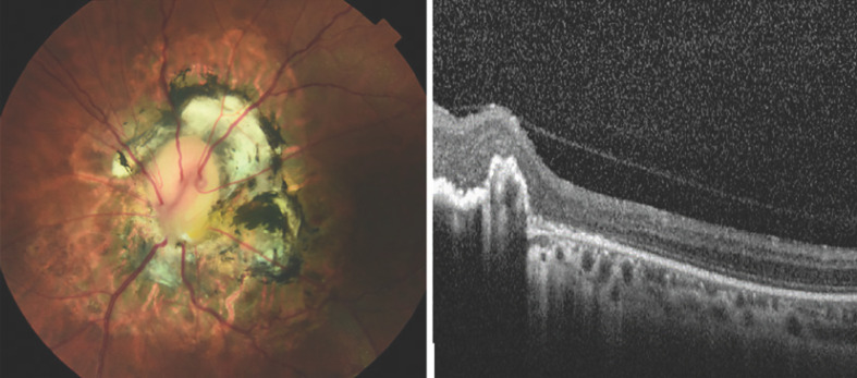
We report a case of morning glory disc anomaly in a young patient with tractional retinal detachment successfully repaired with complex pars plana vitrectomy, membrane peel, laser, and oil tamponade. A 19-year-old female with a history of right morning glory disc anomaly associated with PAX6 gene mutation presented with floaters, photopsia, central scotoma, and visual acuity (VA) of 1/200. A complex macula-involving tractional retinal detachment centered around the optic nerve with a morning glory disc anomaly. Retinal detachment was treated with 25-gauge pars plana vitrectomy with difficult separation of the posterior hyaloid. Fibrous preretinal membranes were peeled, a temporal relaxing retinotomy was required, subretinal fluid was drained through a superonasal retinotomy during air-fluid exchange, endolaser was applied, and tamponade was achieved with 1,000-centistoke silicone oil. The retina remained attached at 1-year follow-up, with VA count fingers throughout. Morning glory disc is a rare congenital anomaly associated with PAX6 gene mutation that most often occurs unilaterally. It is rarely associated with tractional retinal detachment. Optimization of visual outcome is imperative despite a poor visual prognosis.
Keywords: Morning glory disc anomaly; PAX6 gene mutation; Retinal detachment.
Copyright © 2021 by S. Karger AG, Basel.
Conflict of interest statement
The authors have no financial disclosures.
Figures


Similar articles
-
Progress in the Management of Retinal Detachment Associated With Morning Glory Syndrome.Clin Ophthalmol. 2025 Feb 11;19:459-468. doi: 10.2147/OPTH.S505086. eCollection 2025. Clin Ophthalmol. 2025. PMID: 39959883 Free PMC article. Review.
-
MANAGEMENT OF RETINAL DETACHMENT ASSOCIATED WITH MORNING GLORY SYNDROME USING THE HUMAN AMNIOTIC MEMBRANE.Retin Cases Brief Rep. 2024 Jan 1;18(1):18-23. doi: 10.1097/ICB.0000000000001303. Retin Cases Brief Rep. 2024. PMID: 35944558
-
Case Report: Fibroglial Retinal Tissue in Contractile Morning Glory Disc Anomaly.Case Rep Ophthalmol. 2021 Jun 11;12(2):525-530. doi: 10.1159/000510958. eCollection 2021 May-Aug. Case Rep Ophthalmol. 2021. PMID: 34248586 Free PMC article.
-
Outcomes of vitreoretinal surgery in retinal detachment associated with morning glory disc anomaly.Indian J Ophthalmol. 2021 Aug;69(8):2116-2121. doi: 10.4103/ijo.IJO_189_21. Indian J Ophthalmol. 2021. PMID: 34304189 Free PMC article.
-
Postoperative follow-up of a case of atypical morning glory syndrome associated with persistent fetal vasculature.BMC Ophthalmol. 2019 Jul 16;19(1):150. doi: 10.1186/s12886-019-1154-6. BMC Ophthalmol. 2019. PMID: 31311513 Free PMC article. Review.
Cited by
-
Progress in the Management of Retinal Detachment Associated With Morning Glory Syndrome.Clin Ophthalmol. 2025 Feb 11;19:459-468. doi: 10.2147/OPTH.S505086. eCollection 2025. Clin Ophthalmol. 2025. PMID: 39959883 Free PMC article. Review.
-
Vitrectomy Combined With Gas Tamponade for the Treatment of Morning Glory Syndrome With Rhegmatogenous Retinal Detachment: A Case Report.Cureus. 2025 Feb 5;17(2):e78550. doi: 10.7759/cureus.78550. eCollection 2025 Feb. Cureus. 2025. PMID: 40062134 Free PMC article.
References
-
- Kindler P. Morning glory syndrome: unusual congenital optic disk anomaly. Am J Ophthalmol. 1970 Mar;69((3)):376–84. - PubMed
-
- Steinkuller PG. The morning glory disk anomaly: case report and literature review. J Pediatr Ophthalmol Strabismus. 1980 Mar-Apr;17((2)):81–7. - PubMed
-
- Beyer WB, Quencer RM, Osher RH, Morning glory syndrome. A functional analysis including fluorescein angiography, ultrasonography, and computerized tomography. Ophthalmology. 1982 Dec;89((12)):1362–7. - PubMed
-
- Cennamo G, de Crecchio G, Iaccarino G, Forte R. Evaluation of morning glory syndrome with spectral optical coherence tomography and echography. Ophthalmology. 2010 Jun;117((6)):1269–73. - PubMed
Publication types
LinkOut - more resources
Full Text Sources
Miscellaneous
