Case Reports
. 2021 May 27;12(2):464-472.
doi: 10.1159/000512285. eCollection 2021 May-Aug.

Postoperative Macular Proliferative Vitreoretinopathy: A Case Series and Literature Review

Affiliations
Case Reports

Postoperative Macular Proliferative Vitreoretinopathy: A Case Series and Literature Review

Samer Khateb et al. Case Rep Ophthalmol. .

Abstract

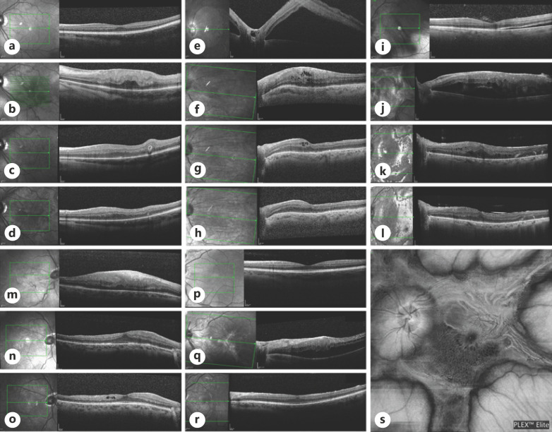
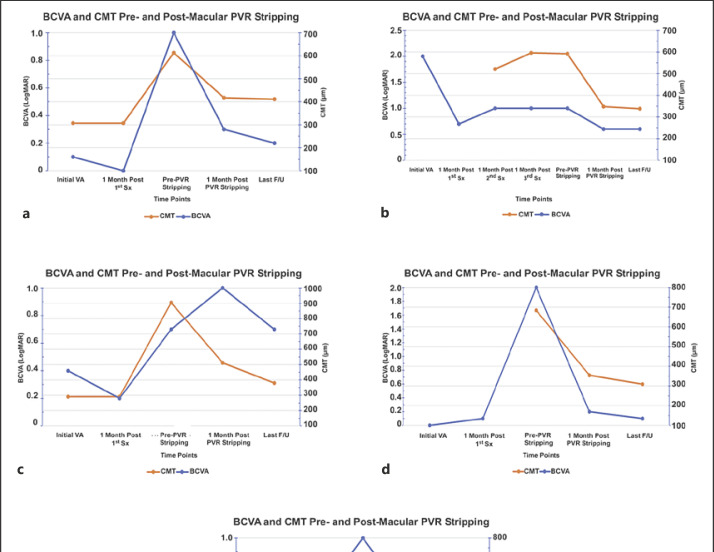
Premacular membranes developing following pars plana vitrectomy (PPV) can cause significant anatomical and functional deficits to the macula. Recent reports showed that postoperative premacular membranes are a localized presentation of macular proliferative vitreoretinopathy (mPVR). Here, we report retrospectively a case series of 5 patients with severe mPVR which developed following uneventful PPV and were followed up to 32 months in the Department of Ophthalmology, Hadassah-Hebrew University Medical Center, Jerusalem, between October 2016 and February 2020. All patients underwent primary repair of rhegmatogenous retinal detachment (RRD) before mPVR developed. Mean best-corrected visual acuity (BCVA) at presentation was 20/76 Snellen (0.58 LogMAR). Median duration of the retinal detachment time until surgery was 1.5 days (range 1-21 days). Mean interval time from last normal follow-up exam to diagnosis of mPVR was 19 days (range 10-28). BCVA dropped from a mean of 20/38 Snellen (0.28 LogMAR) prior to mPVR development to 20/166 Snellen (0.92 LogMAR) following its development, recovering to 20/57 Snellen (0.45 LogMAR) after peeling of membranes. Mean central macular thickness measured by optical coherence tomography decreased from 711 to 354 μm postsurgery. In conclusion, short-term mPVR is a different entity from macular pucker in terms of rapid development, structural distortion, and visual compromise. Surgical treatment significantly restores macular function and anatomy.

Keywords: Epiretinal membrane; Macular proliferative vitreoretinopathy; Macular pucker; Post-operative; Premacular membrane.


Conflict of interest statement

The authors have no conflicts of interest.

Figures

Fig. 1
Fig. 1
Horizontal cross-sections and en face scan of SD-OCT of the macular area for the 5 patients included in the case series: Patient I (a–d), patient II (e–h), patient III (i–l, s), patient IV (m–o), and patient V (p–r). Horizontal OCT cross-sections demonstrate macula-on RRD at presentation (a), developing mPVR leading to macular thickening and disruption of the inner retinal layers (b). Panels (c, d) show restoration of the macular structure following membranes peeling 1 month and in the last follow-up visits, respectively. Patient II presented with macula-off RRD initially (e) followed by mPVR development after 2 PPV surgeries (f). Scans (g, h) show decrease in the macular thickening and restoration of the retinal layers 1 month and as seen in the last follow-up visits post-peeling. Patient III presented with split macula RRD (i), underwent surgical repair followed by severe mPVR (j) seen as macular thickening, hypo-reflective intraretinal cysts, and thick hyper-reflective premacular fibrotic tissue. Surgical repair led to remodeling of the foveal structure as observed 1 month (k) and in the last follow-up visit (l). Patient IV had Macula-on RRD (no scan at presentation) and developed thick mPVR membrane disrupting the inner retinal layers 6 weeks later (m), which resolved almost completely 1 month (n) and 5 months (o) post-op. s En face scan of the macula of patient IV demonstrating unique star-like shape of the PVR membrane encroaching on the posterior pole. Patient V presented macula-on RRD (p) in his LE underwent SB followed by PPV. Three weeks later presented with mPVR (q) causing thickening of the macula without hydration. Membranes peeling restored the macular structure as seen 7 weeks later (r). PVR, proliferative vitreoretinopathy; BCVA, best-corrected visual acuity; SD-OCT, spectral-domain optical coherence tomography; PPV, pars plana vitrectomy; RRD, rhegmatogenous retinal detachment; mPVR, macular proliferative vitreoretinopathy; LE, left eye.
Fig. 2
Fig. 2
BCVA and CMT progression at critical time points of the PVR development and removal. BCVA is presented in LogMAR (blue line) and CMT in µm (orange line). a–e represents patients I–V, respectively. Overall, some inverse correlation between BCVA and CMT was observed, indicating to some extent the importance of membrane stripping in order to improve BCVA. PVR, proliferative vitreoretinopathy; BCVA, best-corrected visual acuity; CMT, central macular thickness.

Similar articles

Cited by

References

    1. Jones MA, Bhide S, Chin E, Ng BG, Rhodenizer D, Zhang VW, et al. Targeted polymerase chain reaction-based enrichment and next generation sequencing for diagnostic testing of congenital disorders of glycosylation. Genet Med. 2011;13((11)):921–32. - PMC - PubMed
    1. Sigler EJ, Randolph JC, Calzada JI. Comparison of morphologic features of macular proliferative vitreoretinopathy and idiopathic epimacular membrane. Retina. 2014;34((8)):1651–7. - PubMed
    1. Guenther SR, Schumann RG, Hagenau F, Wolf A, Priglinger SG, Vogt D. Comparison of surgically excised premacular membranes in eyes with macular pucker and proliferative vitreoretinopathy. Curr Eye Res. 2019;44((3)):341–9. - PubMed
    1. Pastor JC, De La Rúa ER, Martín F. Proliferative vitreoretinopathy: risk factors and pathobiology. Prog Retin Eye Res. 2002;21:127–44. - PubMed
    1. Michels RG. A clinical and histopathologic study of epiretinal membranes affecting the macula and removed by vitreous surgery. Trans Am Ophthalmol Soc. 1982;80:580–656. - PMC - PubMed

Publication types