Hydrogen Sulfide Attenuated Sepsis-Induced Myocardial Dysfunction Through TLR4 Pathway and Endoplasmic Reticulum Stress
- PMID: 34177611
- PMCID: PMC8220204
- DOI: 10.3389/fphys.2021.653601
Hydrogen Sulfide Attenuated Sepsis-Induced Myocardial Dysfunction Through TLR4 Pathway and Endoplasmic Reticulum Stress
Abstract
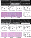
Aims: We examined the change in endogenous hydrogen sulfide (H2S) production and its role in sepsis-induced myocardial dysfunction (SIMD). Results: Significant elevations in plasma cardiac troponin I (cTnI), creatine kinase (CK), tumor necrosis factor-α (TNF-α), and interleukin-1β (IL-1β) were noted in SIMD patients, whereas left ventricular ejection fraction (LVEF), left ventricular fractional shortening (LVFS), and plasma H2S were significantly decreased relative to those in the controls. Plasma H2S was linearly related to LVEF and LVFS. Subsequently, an SIMD model was developed in mice by injecting lipopolysaccharide (LPS), and NaHS, an H2S donor, was used to elucidate the pathophysiological role of H2S. The mice showed decreased ventricular function and increased levels of TNF-α, IL-1β, cTnI, and CK after LPS injections. Toll-like receptor (TLR) 4 protein and endoplasmic reticulum stress (ERS) proteins were over expressed in the SIMD mice. All of the parameters above showed more noticeable variations in cystathionine γ-lyase knockout mice relative to those in wild type mice. The administration of NaHS could improve ventricular function and attenuate inflammation and ERS in the heart. Conclusion: Overall, these findings indicated that endogenous H2S deficiency contributed to SIMD and exogenous H2S ameliorated sepsis-induced myocardial dysfunction by suppressing inflammation and ERS via inhibition of the TLR4 pathway.
Keywords: Toll-like receptor 4; endoplasmic reticulum stress; hydrogen sulfide; myocardial dysfunction; sepsis.
Copyright © 2021 Chen, Teng, Hu, Tian, Jin and Wu.
Conflict of interest statement
The authors declare that the research was conducted in the absence of any commercial or financial relationships that could be construed as a potential conflict of interest.
Figures


Similar articles
-
Hydrogen sulfide alleviates lipopolysaccharide-induced myocardial injury through TLR4-NLRP3 pathway.Physiol Res. 2023 Mar 8;72(1):15-25. doi: 10.33549/physiolres.934928. Epub 2022 Dec 22. Physiol Res. 2023. PMID: 36545872 Free PMC article.
-
[Hydrogen Sulfide Ameliorates Myocardial Injury Caused by Sepsis Through Suppressing ROS-Mediated Endoplasmic Reticulum Stress].Sichuan Da Xue Xue Bao Yi Xue Ban. 2022 Sep;53(5):798-804. doi: 10.12182/20220960106. Sichuan Da Xue Xue Bao Yi Xue Ban. 2022. PMID: 36224681 Free PMC article. Chinese.
-
Significance of hydrogen sulfide in sepsis-induced myocardial injury in rats.Exp Ther Med. 2017 Sep;14(3):2153-2161. doi: 10.3892/etm.2017.4742. Epub 2017 Jul 9. Exp Ther Med. 2017. PMID: 28962136 Free PMC article.
-
Role of hydrogen sulfide in secondary neuronal injury.Neurochem Int. 2014 Jan;64:37-47. doi: 10.1016/j.neuint.2013.11.002. Epub 2013 Nov 14. Neurochem Int. 2014. PMID: 24239876 Review.
-
Research Progress on Mechanisms and Treatment of Sepsis-Induced Myocardial Dysfunction.Int J Gen Med. 2024 Aug 5;17:3387-3393. doi: 10.2147/IJGM.S472846. eCollection 2024. Int J Gen Med. 2024. PMID: 39130486 Free PMC article. Review.
Cited by
-
Exogenous hydrogen sulfide improves non-alcoholic fatty liver disease by inhibiting endoplasmic reticulum stress/NLRP3 inflammasome pathway.Mol Cell Biochem. 2025 Jun;480(6):3813-3839. doi: 10.1007/s11010-025-05220-3. Epub 2025 Feb 8. Mol Cell Biochem. 2025. PMID: 39921790
-
Clinical implications of septic cardiomyopathy: A narrative review.Medicine (Baltimore). 2024 Apr 26;103(17):e37940. doi: 10.1097/MD.0000000000037940. Medicine (Baltimore). 2024. PMID: 38669408 Free PMC article. Review.
-
Mineralocorticoid Receptor Antagonism Prevents the Synergistic Effect of Metabolic Challenge and Chronic Kidney Disease on Renal Fibrosis and Inflammation in Mice.Front Physiol. 2022 Apr 7;13:859812. doi: 10.3389/fphys.2022.859812. eCollection 2022. Front Physiol. 2022. PMID: 35464084 Free PMC article.
-
Unveiling the anti-inflammatory mechanism of exogenous hydrogen sulfide in Kawasaki disease based on network pharmacology and experimental validation.Sci Rep. 2025 Mar 3;15(1):7410. doi: 10.1038/s41598-025-91998-7. Sci Rep. 2025. PMID: 40033067 Free PMC article.
-
Determination of sulfide in complex biofilm matrices using silver-coated, 4-mercaptobenzonitrile-modified gold nanoparticles, encapsulated in ZIF-8 as surface-enhanced Raman scattering nanoprobe.Mikrochim Acta. 2023 Nov 22;190(12):475. doi: 10.1007/s00604-023-06071-9. Mikrochim Acta. 2023. PMID: 37991569
References
LinkOut - more resources
Full Text Sources
Research Materials

