

Regenerated Microvascular Networks in Ischemic Skeletal Muscle
- PMID: 34177614
- PMCID: PMC8231913
- DOI: 10.3389/fphys.2021.662073
Regenerated Microvascular Networks in Ischemic Skeletal Muscle
Abstract
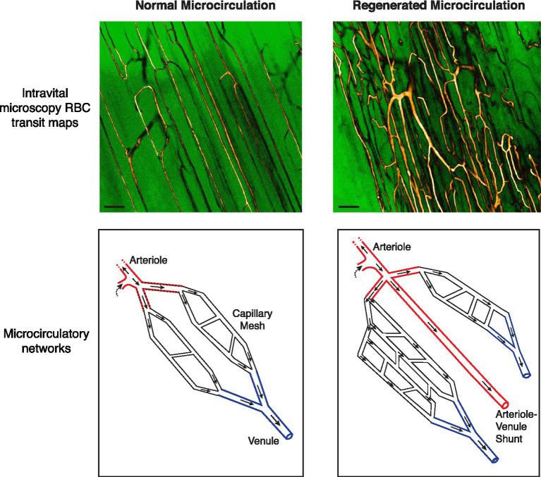
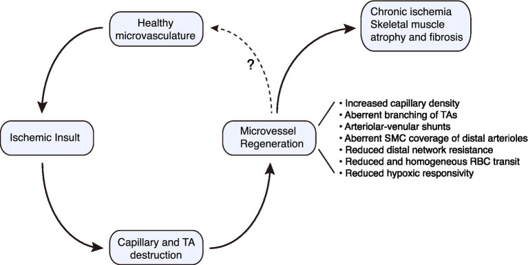
Skeletal muscle is the largest organ in humans. The viability and performance of this metabolically demanding organ are exquisitely dependent on the integrity of its microcirculation. The architectural and functional attributes of the skeletal muscle microvasculature are acquired during embryonic and early postnatal development. However, peripheral vascular disease in the adult can damage the distal microvasculature, together with damaging the skeletal myofibers. Importantly, adult skeletal muscle has the capacity to regenerate. Understanding the extent to which the microvascular network also reforms, and acquires structural and functional competence, will thus be critical to regenerative medicine efforts for those with peripheral artery disease (PAD). Herein, we discuss recent advances in studying the regenerating microvasculature in the mouse hindlimb following severe ischemic injury. We highlight new insights arising from real-time imaging of the microcirculation. This includes identifying otherwise hidden flaws in both network microarchitecture and function, deficiencies that could underlie the progressive nature of PAD and its refractoriness to therapy. Recognizing and overcoming these vulnerabilities in regenerative angiogenesis will be important for advancing treatment options for PAD.
Keywords: angiogenesis; intravital microscopy; peripheral artery disease; skeletal muscle; smooth muscle cell.
Copyright © 2021 Yin, Arpino, Lee and Pickering.
Conflict of interest statement
The authors declare that the research was conducted in the absence of any commercial or financial relationships that could be construed as a potential conflict of interest.
Figures
Similar articles
-
Four-Dimensional Microvascular Analysis Reveals That Regenerative Angiogenesis in Ischemic Muscle Produces a Flawed Microcirculation.Circ Res. 2017 Apr 28;120(9):1453-1465. doi: 10.1161/CIRCRESAHA.116.310535. Epub 2017 Feb 7. Circ Res. 2017. PMID: 28174322
-
Recovery of blood flow regulation in microvascular resistance networks during regeneration of mouse gluteus maximus muscle.J Physiol. 2019 Mar;597(5):1401-1417. doi: 10.1113/JP277247. Epub 2019 Feb 3. J Physiol. 2019. PMID: 30575953 Free PMC article.
-
A novel approach for comparative study of periosteum, muscle, subcutis, and skin microcirculation by intravital fluorescence microscopy.Microvasc Res. 1998 Jul;56(1):30-42. doi: 10.1006/mvre.1998.2077. Microvasc Res. 1998. PMID: 9683561
-
Systematic Interrogation of Angiogenesis in the Ischemic Mouse Hind Limb: Vulnerabilities and Quality Assurance.Arterioscler Thromb Vasc Biol. 2020 Oct;40(10):2454-2467. doi: 10.1161/ATVBAHA.120.315028. Epub 2020 Aug 13. Arterioscler Thromb Vasc Biol. 2020. PMID: 32787524 Free PMC article.
-
Pathophysiology of Intermittent Claudication in Peripheral Artery Disease.Circ J. 2017 Feb 24;81(3):281-289. doi: 10.1253/circj.CJ-16-1286. Epub 2017 Jan 26. Circ J. 2017. PMID: 28123169 Review.
Cited by
-
Interdependence of Angiogenesis and Arteriogenesis in Development and Disease.Int J Mol Sci. 2022 Mar 31;23(7):3879. doi: 10.3390/ijms23073879. Int J Mol Sci. 2022. PMID: 35409246 Free PMC article. Review.
-
Endothelial-mesenchymal transition in skeletal muscle: Opportunities and challenges from 3D microphysiological systems.Bioeng Transl Med. 2024 Jan 29;9(5):e10644. doi: 10.1002/btm2.10644. eCollection 2024 Sep. Bioeng Transl Med. 2024. PMID: 39553431 Free PMC article. Review.
-
Multi-modality imaging for assessment of the microcirculation in peripheral artery disease: Bench to clinical practice.Am Heart J Plus. 2024 May 8;42:100400. doi: 10.1016/j.ahjo.2024.100400. eCollection 2024 Jun. Am Heart J Plus. 2024. PMID: 38779485 Free PMC article.
References
-
- Aboyans V., Ricco J. B., Bartelink M. E. L., Bjorck M., Brodmann M., Cohnert T., et al. . (2018). 2017 ESC guidelines on the diagnosis and treatment of peripheral arterial diseases, in collaboration with the European Society for Vascular Surgery (ESVS): document covering atherosclerotic disease of extracranial carotid and vertebral, mesenteric, renal, upper and lower extremity arteries. Endorsed by: the European stroke organization (ESO) The task force for the diagnosis and treatment of peripheral arterial diseases of the European Society of Cardiology (ESC) and of the European Society for Vascular Surgery (ESVS). Eur. Heart J. 39, 763–816. 10.1093/eurheartj/ehx095, PMID: - DOI - PubMed
LinkOut - more resources
Full Text Sources