. 2021 Jun 10:12:613630.
doi: 10.3389/fneur.2021.613630. eCollection 2021.

Functional Hyperconnectivity and Task-Based Activity Changes Associated With Neuropathic Pain After Spinal Cord Injury: A Pilot Study

Affiliations

Functional Hyperconnectivity and Task-Based Activity Changes Associated With Neuropathic Pain After Spinal Cord Injury: A Pilot Study

Shana R Black et al. Front Neurol. .

Abstract

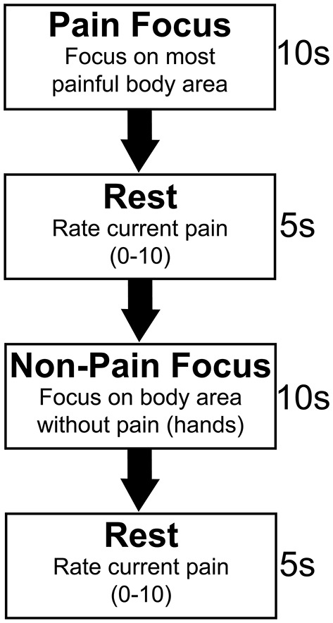
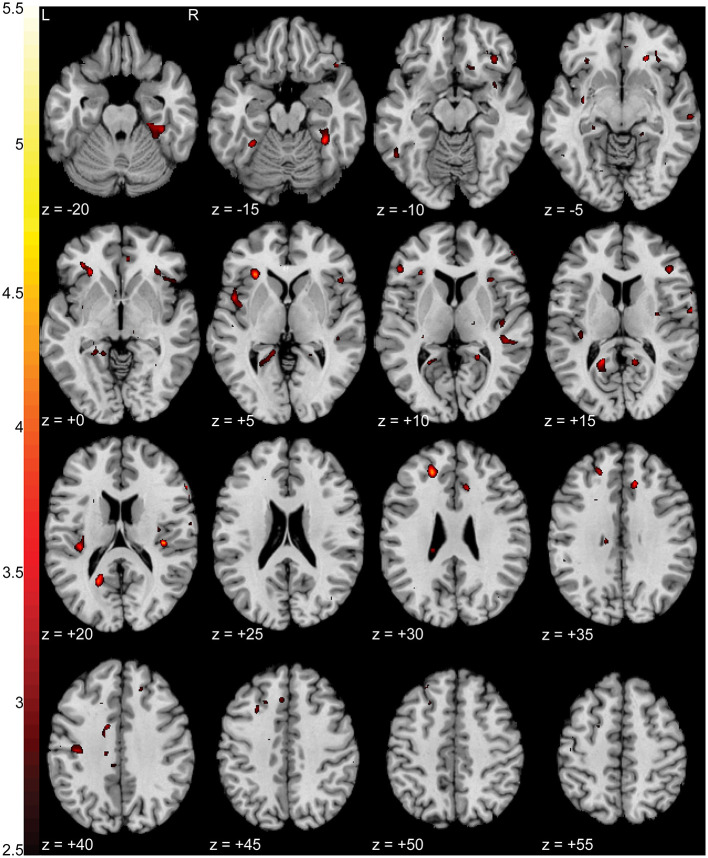
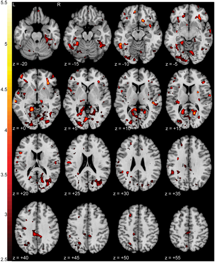
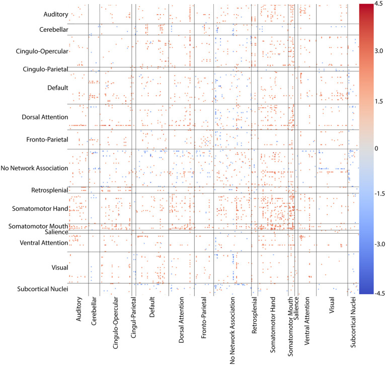
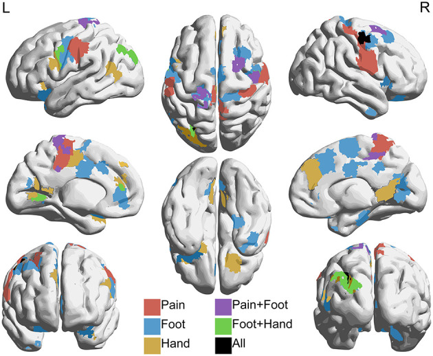
Neuropathic pain (NP) is a devastating chronic pain condition affecting roughly 80% of the spinal cord injury (SCI) patient population. Current treatment options are largely ineffective and neurophysiological mechanisms of NP are not well-understood. Recent studies in neuroimaging have suggested that NP patients have differential patterns of functional activity that are dependent upon the neurological condition causing NP. We conducted an exploratory pilot study to examine functional activation and connectivity in SCI patients with chronic NP compared to SCI patients without NP. We developed a novel somatosensory attention task to identify short term fluctuations in neural activity related to NP vs. non-painful somatosensation using functional magnetic resonance imaging (fMRI). We also collected high-resolution resting state fMRI to identify connectivity-based correlations over time between the two groups. We observed increased activation during focus on NP in brain regions associated with somatosensory integration and representational knowledge in pain subjects when compared with controls. Similarly, NP subjects showed increased connectivity at rest in many of the same areas of the brain, with positive correlations between somatomotor networks, the dorsal attention network, and regions associated with pain and specific areas of painful and non-painful sensation within our cohort. Although this pilot analysis did not identify statistically significant differences between groups after correction for multiple comparisons, the observed correlations between NP and functional activation and connectivity align with a priori hypotheses regarding pain, and provide a well-controlled preliminary basis for future research in this severely understudied patient population. Altogether, this study presents a novel task, identifies regions of increased task-based activation associated with NP after SCI in the insula, prefrontal, and medial inferior parietal cortices, and identifies similar regions of increased functional connectivity associated with NP after SCI in sensorimotor, cingulate, prefrontal, and inferior medial parietal cortices. This, along with our complementary results from a structurally based analysis, provide multi-modal evidence for regions of the brain specific to the SCI cohort as novel areas for further study and potential therapeutic targeting to improve outcomes for NP patients.

Keywords: fMRI; functional connectivity; functional magnetic resonance imaging; neuropathic pain; resting-state fMRI; spinal cord injury; task-based fMRI.


Conflict of interest statement

The authors declare that the research was conducted in the absence of any commercial or financial relationships that could be construed as a potential conflict of interest.

Figures

Figure 1
Figure 1
Somatosensory focus task paradigm. Pain subjects were asked to alternate between two active focus periods of 10 s. For the first focus period, the pain focus, subjects were asked to focus only on their most painful area. Subjects were asked to focus on their intrinsic somatosensation in a body area in which they did not have pain symptoms for the second, non-pain focus period. This was the hands for all subjects. Five second rest periods separated focus periods during which time pain subjects verbally rated their pain on a 0–10 rating scale. Control subjects were asked to focus on the intrinsic somatosensation in the same two body areas as their age, sex, and injury level matched pain group counterpart.
Figure 2
Figure 2
Individually defined association maps used as ROIs for connectivity analysis. Association maps isolated with Neurosynth (neurosynth.org) using “pain” (A), “foot” (B), and “hand” (C) as individual keywords. The foot and hand keywords were chosen as correlates for painful and non-painful somatosensory activity, respectively. Selected axial (top row), sagittal (bottom row, left), and coronal (bottom row, right) slices representing the spatial distribution of each functional association area are shown for each keyword. Each association map was treated as an individual ROI for inclusion in our connectivity analysis.
Figure 3
Figure 3
Flow diagram of subjects enrolled and included in resting state connectivity (rs-FMRI) and task-based (t-fMRI) analyses.
Figure 4
Figure 4
Increased activation in NP subjects compared to controls during the pain focus state in contrast to the non-pain focus state. T-statistic mapping of increased BOLD activation (p < 0.05 uncorrected) in NP subjects during the pain focus state when compared to the pain minus non-pain contrast in control subjects. Increased activity was seen in bilateral anterior and posterior insula, left retrosplenial cortex, and ventromedial prefrontal cortex. Clusters with fewer than 10 contiguous voxels were filtered for clarity. The difference between axial slices is 5 mm and z-values are relative to the AC/PC plane.
Figure 5
Figure 5
Increased activation in NP subjects during the pain focus state compared to the non-pain focus state. T-statistic mapping of increased BOLD activation (p < 0.05 uncorrected) during the pain focus state when compared to the non-pain focus state in the NP subject group. Increased activity was seen bilaterally in precuneus, anterior insula, middle temporal gyrus, middle cingulate cortex, and orbitofrontal cortex, and unilaterally in the left lingual gyrus. Clusters fewer than 10 contiguous voxels were filtered for clarity. The difference between axial slices is 5 mm and z-values are relative to the AC/PC plane.
Figure 6
Figure 6
Connectivity matrix of regions with altered connectivity between NP and control groups. T-statistic values for whole-brain, all-to-all comparisons between groups for the 361 ROI combined cortical, subcortical, and cerebellar parcellation. Somatomotor hand, somatomotor mouth, and dorsal attention networks showed widespread increases in connectivity with nearly every other network. Hyperconnectivity was particularly dense in somatomotor-somatomotor connections. Only connections with differences at p < 0.05 uncorrected are colored. Warm colors (reds) indicate an increase in connectivity in the NP group when compared to controls. Cool colors (blues) indicate a decrease in the NP group.
Figure 7
Figure 7
Regions with increased connectivity to pain, foot, and hand association regions in NP subjects compared to controls. Regions within the 361 combined parcellation showing increased connectivity (p < 0.05 uncorrected) with individually defined regions associated with pain, foot, and hand in NP subjects when compared to controls. Hyperconnectivity was seen between the pain region and parts of the dorsal attention, ventral attention, and somatomotor networks. The foot region showed increased connectivity with parts of the cingulo-opercular, dorsal attention, fronto-parietal, and somatomotor networks. The hand region showed increased connectivity with default, dorsal attention, fronto-parietal, ventral attention, and somatomotor networks. Lateral (top left and right), medial (middle left and right), anterior (bottom left), posterior (bottom right), superior (top middle), and inferior (bottom middle) views are shown.

References

    1. Finnerup NB, Haroutounian S, Kamerman P, Baron R, Bennett DLH, Bouhassira D, et al. . Neuropathic pain: an updated grading system for research and clinical practice. Pain. (2016) 157:1599–606. 10.1097/j.pain.0000000000000492 - DOI - PMC - PubMed
    1. Burke D, Fullen BM, Stokes D, Lennon O. Neuropathic pain prevalence following spinal cord injury: a systematic review and meta-analysis. Eur J Pain. (2017) 21:29–44. 10.1002/ejp.905 - DOI - PubMed
    1. Center NSCIS . Spinal Cord Injury Facts and Figures at a Glance 2020 SCI Data Sheet. (2020). Available online at: www.msktc.org/sci/model-system-centers (accessed July 2, 2020).
    1. Finnerup NB, Baastrup C, Jensen TS. Neuropathic pain following spinal cord injury pain: mechanisms and treatment. Scand J Pain. (2009) 1(Suppl. 1):S3–11. 10.1016/S1877-8860(09)70003-5 - DOI
    1. Zheng W, Chen Q, Chen X, Wan L, Qin W, Qi Z, et al. . Brain white matter impairment in patients with spinal cord injury. Neural Plast. (2017) 2017:1–8. 10.1155/2017/4671607 - DOI - PMC - PubMed