

Contribution of CT-Scan Analysis by Artificial Intelligence to the Clinical Care of TBI Patients
- PMID: 34177773
- PMCID: PMC8222716
- DOI: 10.3389/fneur.2021.666875
Contribution of CT-Scan Analysis by Artificial Intelligence to the Clinical Care of TBI Patients
Abstract
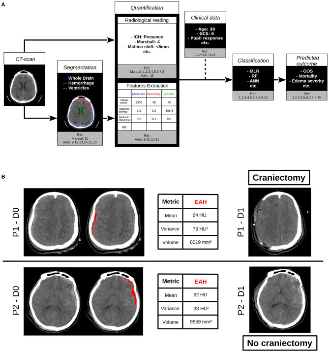
The gold standard to diagnose intracerebral lesions after traumatic brain injury (TBI) is computed tomography (CT) scan, and due to its accessibility and improved quality of images, the global burden of CT scan for TBI patients is increasing. The recent developments of automated determination of traumatic brain lesions and medical-decision process using artificial intelligence (AI) represent opportunities to help clinicians in screening more patients, identifying the nature and volume of lesions and estimating the patient outcome. This short review will summarize what is ongoing with the use of AI and CT scan for patients with TBI.
Keywords: artificial intelligence; classification; computed tomography; review; segmentation; traumatic brain injury.
Copyright © 2021 Brossard, Lemasson, Attyé, de Busschère, Payen, Barbier, Grèze and Bouzat.
Conflict of interest statement
The authors declare that the research was conducted in the absence of any commercial or financial relationships that could be construed as a potential conflict of interest.
Figures
Similar articles
-
Radiological features of brain hemorrhage through automated segmentation from computed tomography in stroke and traumatic brain injury.Front Neurol. 2023 Sep 28;14:1244672. doi: 10.3389/fneur.2023.1244672. eCollection 2023. Front Neurol. 2023. PMID: 37840934 Free PMC article.
-
Automated identification and quantification of traumatic brain injury from CT scans: Are we there yet?Medicine (Baltimore). 2022 Nov 25;101(47):e31848. doi: 10.1097/MD.0000000000031848. Medicine (Baltimore). 2022. PMID: 36451512 Free PMC article.
-
Automated Outcome Classification of Computed Tomography Imaging Reports for Pediatric Traumatic Brain Injury.Acad Emerg Med. 2016 Feb;23(2):171-8. doi: 10.1111/acem.12859. Epub 2016 Jan 14. Acad Emerg Med. 2016. PMID: 26766600 Free PMC article.
-
The effect of scan and patient parameters on the diagnostic performance of AI for detecting coronary stenosis on coronary CT angiography.Clin Imaging. 2022 Apr;84:149-158. doi: 10.1016/j.clinimag.2022.01.016. Epub 2022 Feb 3. Clin Imaging. 2022. PMID: 35217284
-
Artificial Intelligence and Machine Learning in Nuclear Medicine: Future Perspectives.Semin Nucl Med. 2021 Mar;51(2):170-177. doi: 10.1053/j.semnuclmed.2020.08.003. Epub 2020 Sep 12. Semin Nucl Med. 2021. PMID: 33509373 Review.
Cited by
-
The application value of CT radiomics features in predicting pressure amplitude correlation index in patients with severe traumatic brain injury.Front Neurol. 2022 Aug 25;13:905655. doi: 10.3389/fneur.2022.905655. eCollection 2022. Front Neurol. 2022. PMID: 36090879 Free PMC article.
-
The Use of Artificial Intelligence in the Liver Histopathology Field: A Systematic Review.Diagnostics (Basel). 2024 Feb 10;14(4):388. doi: 10.3390/diagnostics14040388. Diagnostics (Basel). 2024. PMID: 38396427 Free PMC article. Review.
-
Prediction of therapeutic intensity level from automatic multiclass segmentation of traumatic brain injury lesions on CT-scans.Sci Rep. 2023 Nov 17;13(1):20155. doi: 10.1038/s41598-023-46945-9. Sci Rep. 2023. PMID: 37978266 Free PMC article. Clinical Trial.
-
Henry gas solubility optimization double machine learning classifier for neurosurgical patients.PLoS One. 2023 May 11;18(5):e0285455. doi: 10.1371/journal.pone.0285455. eCollection 2023. PLoS One. 2023. PMID: 37167226 Free PMC article.
-
AI-Based Decision Support System for Traumatic Brain Injury: A Survey.Diagnostics (Basel). 2023 May 5;13(9):1640. doi: 10.3390/diagnostics13091640. Diagnostics (Basel). 2023. PMID: 37175031 Free PMC article. Review.
References
Publication types
LinkOut - more resources
Full Text Sources