

Investigation Into the Predictive Potential of Three-Dimensional Ultrasonographic Placental Volume and Vascular Indices in Gestational Diabetes Mellitus
- PMID: 34177812
- PMCID: PMC8222907
- DOI: 10.3389/fendo.2021.689888
Investigation Into the Predictive Potential of Three-Dimensional Ultrasonographic Placental Volume and Vascular Indices in Gestational Diabetes Mellitus
Abstract
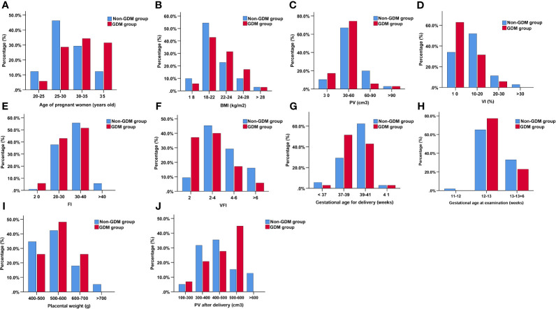
Background: The use of ultrasonography in pregnancies complicated with gestational diabetes mellitus (GDM) can vary according to clinical practice. This study aims to compare the changes of placental volume (PV) and vascular indices measured by three-dimensional (3D) Power Doppler between pregnant women with and without GDM.
Materials and methods: This was a prospective study of singleton pregnancies who took the early nuchal translucency examination from January 2018 to September 2019. Data on PV and vascular indices including vascularization index (VI), flow index (FI), and vascularization flow index (VFI) between pregnant women with and without GDM were measured by 3D Power Doppler ultrasound machine. Univariate and multivariate logistic regression determined the association between risk factors and GDM. Receiver operating characteristic (ROC) and area under the ROC curve (AUC) were applied to evaluate the diagnostic value of different parameters for GDM.
Results: Of the 141 pregnant women enrolled, 35 developed GDM and 106 did not. The maternal age and gravida in the GDM group were significantly higher than that in the non-GDM group. The PV, VI, FI, and VFI in the GDM group were significantly lower than that in the non-GDM group. There were no significant differences in other clinical parameters between the two groups. After adjustments in multivariate logistic regression analysis, significant differences were observed in VI [odds ratio (OR) = 0.98, 95% confidence interval (CI) = 0.951-1.002], FI (OR = 0.93, 955 CI: 0.86-1.00), and VFI (OR = 0.67, 95% CI = 0.52-0.87). ROC analysis indicated that the combination of maternal age, gravida, PV, and VFI was more accurate as a marker for detecting GDM than the PV, VI, FI, or VFI alone.
Conclusions: The 3D ultrasonography results suggest that PV and vascular indices (VI, FI, and VFI) during the first trimester may serve as potential markers for GDM diagnosis. The combination of maternal age, gravida, and sonographic markers may have good diagnostic values for GDM, which should be confirmed by further investigations.
Keywords: flow index; gestational diabetes mellitus; placental volume; three-dimensional power Doppler; vascularization flow index; vascularization index.
Copyright © 2021 Han, Zhang, Li, Chiu, Yin and Hou.
Conflict of interest statement
The authors declare that the research was conducted in the absence of any commercial or financial relationships that could be construed as a potential conflict of interest.
Figures


Similar articles
-
Comparison of placental three-dimensional power Doppler indices and volume in the first and the second trimesters of pregnancy complicated by gestational diabetes mellitus.J Matern Fetal Neonatal Med. 2019 Nov;32(22):3784-3791. doi: 10.1080/14767058.2018.1472226. Epub 2018 Sep 10. J Matern Fetal Neonatal Med. 2019. PMID: 29716432
-
Placental three-dimensional power Doppler indices in mid-pregnancy and late pregnancy complicated by gestational diabetes mellitus.Prenat Diagn. 2013 Oct;33(10):952-8. doi: 10.1002/pd.4172. Epub 2013 Jul 3. Prenat Diagn. 2013. PMID: 23740806
-
Placental volume and vascular flow assessed by 3D power Doppler and adverse pregnancy outcomes.Placenta. 2011 Mar;32(3):230-4. doi: 10.1016/j.placenta.2011.01.010. Epub 2011 Feb 4. Placenta. 2011. PMID: 21295850 Free PMC article.
-
Placental structural abnormalities in gestational diabetes and when they develop: A scoping review.Placenta. 2021 Dec;116:58-66. doi: 10.1016/j.placenta.2021.04.005. Epub 2021 Apr 26. Placenta. 2021. PMID: 33958235 Free PMC article.
-
Available evidence on fetal liver changes detected by ultrasound in pregnant women with gestational diabetes mellitus: Systematic review.J Clin Ultrasound. 2024 Oct;52(8):1121-1128. doi: 10.1002/jcu.23760. Epub 2024 Jul 12. J Clin Ultrasound. 2024. PMID: 38994688
Cited by
-
Reference charts for first-trimester placental volume derived using OxNNet.Ultrasound Obstet Gynecol. 2025 Sep;66(3):337-346. doi: 10.1002/uog.29300. Epub 2025 Aug 1. Ultrasound Obstet Gynecol. 2025. PMID: 40746163 Free PMC article.
-
Predictive value of ultrasonic artificial intelligence in placental characteristics of early pregnancy for gestational diabetes mellitus.Front Endocrinol (Lausanne). 2024 Mar 13;15:1344666. doi: 10.3389/fendo.2024.1344666. eCollection 2024. Front Endocrinol (Lausanne). 2024. PMID: 38544693 Free PMC article.
-
Prediction of Gestational Diabetes Mellitus (GDM) risk in early pregnancy based on clinical data and ultrasound information: a nomogram.BMC Med Inform Decis Mak. 2025 Mar 18;25(1):138. doi: 10.1186/s12911-025-02962-4. BMC Med Inform Decis Mak. 2025. PMID: 40102801 Free PMC article.
-
Influence of gestational diabetes mellitus on the cardiovascular system and its underlying mechanisms.Front Endocrinol (Lausanne). 2025 May 16;16:1474643. doi: 10.3389/fendo.2025.1474643. eCollection 2025. Front Endocrinol (Lausanne). 2025. PMID: 40453589 Free PMC article. Review.
References
MeSH terms
LinkOut - more resources
Full Text Sources