. 2021 Jun 11:12:677634.
doi: 10.3389/fmicb.2021.677634. eCollection 2021.

Porcine RIG-I and MDA5 Signaling CARD Domains Exert Similar Antiviral Function Against Different Viruses

Affiliations

Porcine RIG-I and MDA5 Signaling CARD Domains Exert Similar Antiviral Function Against Different Viruses

Shuangjie Li et al. Front Microbiol. .

Abstract

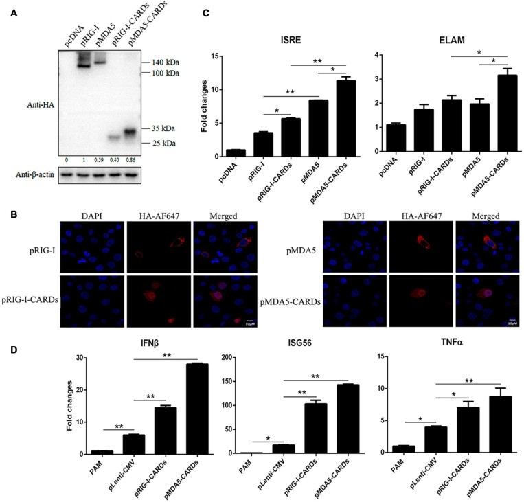
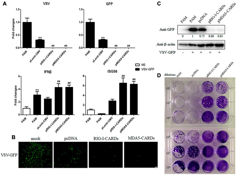
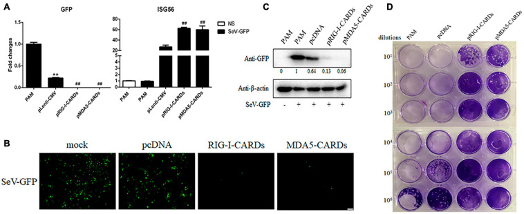
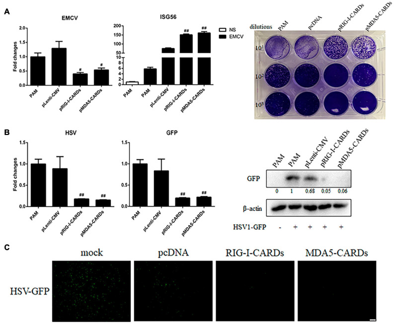
The RIG-I-like receptors (RLRs) RIG-I and MDA5 play critical roles in sensing and fighting viral infections. Although RIG-I and MDA5 have similar molecular structures, these two receptors have distinct features during activation. Further, the signaling domains of the N terminal CARD domains (CARDs) in RIG-I and MDA5 share poor similarity. Therefore, we wonder whether the CARDs of RIG-I and MDA5 play similar roles in signaling and antiviral function. Here we expressed porcine RIG-I and MDA5 CARDs in 293T cells and porcine alveolar macrophages and found that MDA5 CARDs exhibit higher expression and stronger signaling activity than RIG-I CARDs. Nevertheless, both RIG-I and MDA5 CARDs exert comparable antiviral function against several viruses. Transcriptome analysis showed that MDA5 CARDs are more effective in regulating downstream genes. However, in the presence of virus, both RIG-I and MDA5 CARDs exhibit similar effects on downstream gene transcriptions, reflecting their antiviral function.

Keywords: CARD; RLRs; antiviral function; porcine; signaling activity; transcriptome.


Conflict of interest statement

The authors declare that the research was conducted in the absence of any commercial or financial relationships that could be construed as a potential conflict of interest.

Figures

FIGURE 1
FIGURE 1
The protein expression, cellular localization and signaling activity of porcine RIG-I and MDA5 CARDs. (A) The pRIG-I-HA, pMDA5-HA, pRIG-I-CARDs-HA, pMDA5-CARDs-HA, and vector pcDNA (0.5 μg each) were transfected into 293T cells in 24-well plates (3 × 10⁵ cells/well) for 24 h using Lipofectamine 2000 (Thermo Fisher Scientific). The cells samples were detected by Western-blotting with anti-HA and anti-actin mAb. The densitometry values after actin normalization were shown below the blot. (B) PAMs grown on 15-nm glass bottom cell culture dish (5 × 10⁵ cells) were transfected with the indicated pcDNA expression plasmids (0.75 μg each) using TransIT-LT1 Transfection Reagent for 24 h. The cells were examined for localization by con-focal fluorescence microscopy. (C) PAMs grown in 96-well plates (2 × 10⁴ cells/well) were transfected with 20 ng indicated pcDNA expression plasmids, plus ISRE-Fluc/ELAM-Fluc (10 ng) and Rluc (0.2 ng) using TransIT-LT1 Transfection Reagent for 24 h. The luciferase activities were measured with Double-Luciferase Reporter Assay. (D) PAMs grown on 24-well plate (3 × 10⁵ cells/well) were transfected with pLenti-pRIG-I-CARDs-HA and pLenti-pMDA5-CARDs-HA (0.5 μg each) and pLenti-CMV vector using TransIT-LT1 Transfection Reagent for 24 h. Then the cells were analyzed by RT-qPCR for downstream gene expressions as indicated. The signs “*” and “**” denote p < 0.05 and p < 0.01, respectively.
FIGURE 2
FIGURE 2
The anti-VSV activity of porcine RIG-I and MDA5 CARDs. (A) PAMs grown on 24-well plates (3 × 10⁵ cells/well) were transfected with pLenti-pRIG-I-CARDs-HA and pLenti-pMDA5-CARDs-HA and pLenti-CMV vector (0.5 μg each) using TransIT-LT1 Transfection Reagent for 24 h. Then the cells were infected with VSV-GFP virus at MOI of 0.01 for 12 h and analyzed by RT-qPCR for virus replication and downstream gene expressions as indicated. (B–D) PAMs grown on 24-well plate were transfected and infected with VSV as above. The GFP signals were visualized under microscope (B). The cells samples were detected by Western-blotting with anti-GFP mAb, with the densitometry values after actin normalization shown below the GFP blot. (C). The supernatants from VSV infected PAMs were subjected to infection of Vero cells and the viral plaque were observed at 24 h post infection (D). **p < 0.01 vs. blank controls or mock transfection; ##p < 0.01 vs. vector controls.
FIGURE 3
FIGURE 3
The anti-SeV activity of porcine RIG-I and MDA5 CARDs. (A) PAMs grown on 24-well plates (3 × 10⁵ cells/well) were transfected with pLenti-pRIG-I-CARDs-HA and pLenti-pMDA5-CARDs-HA and pLenti-CMV vector (0.5 μg each) for 24 h. Then the cells were infected with SeV-GFP virus at MOI of 0.01 for 12 h and analyzed by RT-qPCR for virus replication and downstream gene expressions as indicated. (B–D) PAMs grown on 24-well plate were transfected and infected as above. The GFP signals were visualized under microscope (B). The cells samples were detected by Western-blotting with anti-GFP mAb, with the densitometry values after actin normalization shown below the GFP blot. (C). The supernatants from SeV infected PAMs were subjected to infection of Vero cells and the viral plaque were visualized at 24 h post infection (D). **p < 0.01 vs. blank controls; ##p < 0.01 vs. vector controls.
FIGURE 4
FIGURE 4
The anti-EMCV and HSV-1 activity of porcine RIG-I and MDA5 CARDs. (A) PAMs grown on 24-well plates (3 × 10⁵ cells/well) were transfected with the indicated CARDs expression plasmids and control vectors (0.5 μg each) using TransIT-LT1 Transfection Reagent for 24 h. Then the cells were infected with EMCV virus at MOI of 0.01 for 12 h and analyzed by RT-qPCR for virus replication, downstream gene expression and by plaque assay for observation of plaque formation in Vero cells at 24 h post infection. (B,C) PAMs grown on 24-well plate (3 × 10⁵ cells/well) were transfected with CARDs expression plasmids and pLenti-CMV vector (0.5 μg each) using TransIT-LT1 Transfection Reagent for 24 h. Then the cells were infected with HSV-1-GFP virus at MOI of 0.01 for 12 h. The cells samples were analyzed by RT-qPCR for virus replication and downstream gene expressions as indicated or detected by Western-blotting with anti-GFP mAb (B). The GFP signals were visualized under microscope (C). #p < 0.05, ##p < 0.01 vs. vector controls.
FIGURE 5
FIGURE 5
The anti-influenza virus H9N2 activity of porcine RIG-I and MDA5 CARDs. PAMs grown on 24-well plates (3 × 10⁵ cells/well) were transfected with CARDs expression plasmids and pLenti-CMV vector (0.5 μg each) using TransIT-LT1 Transfection Reagent for 24 h. Then the cells were infected with H9N2 virus at MOI of 0.01 for 12 h. The cells samples were analyzed by RT-qPCR for virus replication and downstream gene expressions as indicated (A) or detected by Western-blotting using the indicated antibodies (B). The densitometry values after actin normalization were shown below the blots. **p < 0.01 vs. blank controls; ##p < 0.01 vs. vector controls.
FIGURE 6
FIGURE 6
The anti-influenza virus H1N1 activity of porcine RIG-I and MDA5 CARDs. PAMs grown on 24-well plates (3 × 10⁵ cells/well) were transfected with CARDs expression plasmids and pLenti-CMV vector (0.5 μg each) using TransIT-LT1 Transfection Reagent for 24 h. Then the cells were infected with H1N1 virus at MOI of 0.01 for 12 h. The cells samples were analyzed by RT-qPCR for viral M gene and downstream ISG56 gene expressions (A) or detected by Western-blotting using the indicated antibodies (B). The densitometry values after actin normalization were shown below the blots. **p < 0.01 vs. blank controls; #p < 0.05, ##p < 0.01 vs. vector controls.
FIGURE 7
FIGURE 7
The transcriptome analysis of CARDs transfected PAMs with or without H9N2 infection. (A) The heat map of clustered differentially expressed genes (DEGs) in porcine RIG-I and MDA5 transfected PAMs with or without H9N2 infection (0.01 MOI for 12 h). P, PAM mock control; V, pcDNA vector transfection control; R, porcine RIG-I CARDs; M, porcine MDA5 CARDs. The last V denotes H9N2 infection. (B) The Venn’s diagrams of several groups of DEGs. RV, RIG-I CARDs vs. pcDNA control; MV, MDA5 CARDs vs. pcDNA control; RVV, RIG-I-CARDs vs. vector control with H9N2 infection; MVV, MDA5-CARDs vs. vector control with H9N2 infection. (C) The subcluster with the majority DEGs being ISGs, H9N2 infection causes general upregulations of ISGs, whereas two CARDs induce pronounced upregulations of ISGs; upon H9N2 infection (V), the upregulated ISGs go down.

Similar articles

Cited by

References

    1. Brisse M., Ly H. (2019). Comparative structure and function analysis of the RIG-I-like receptors: RIG-I and MDA5. Front. Immunol. 10:1586. 10.3389/fimmu.2019.01586 - DOI - PMC - PubMed
    1. Cao L., Ge X., Gao Y., Herrler G., Ren Y., Ren X., et al. (2015). Porcine epidemic diarrhea virus inhibits dsRNA-induced interferon-beta production in porcine intestinal epithelial cells by blockade of the RIG-I-mediated pathway. Virol. J. 12:127. - PMC - PubMed
    1. Chen N., Xia P., Li S., Zhang T., Wang T. T., Zhu J. (2017). RNA sensors of the innate immune system and their detection of pathogens. IUBMB Life 69 297–304. 10.1002/iub.1625 - DOI - PMC - PubMed
    1. Dong X. Y., Liu W. J., Zhao M. Q., Wang J. Y., Pei J. J., Luo Y. W., et al. (2013). Classical swine fever virus triggers RIG-I and MDA5-dependent signaling pathway to IRF-3 and NF-kappaB activation to promote secretion of interferon and inflammatory cytokines in porcine alveolar macrophages. Virol. J. 10:286. 10.1186/1743-422x-10-286 - DOI - PMC - PubMed
    1. Ge S., Li J., Fan X., Liu F., Li L., Wang Q., et al. (2018). Molecular characterization of African swine fever virus, China, 2018. Emerg. Infect. Dis. 24 2131–2133. - PMC - PubMed

LinkOut - more resources