. 2021 Jun 10:12:690821.
doi: 10.3389/fimmu.2021.690821. eCollection 2021.

Factor D Inhibition Blocks Complement Activation Induced by Mutant Factor B Associated With Atypical Hemolytic Uremic Syndrome and Membranoproliferative Glomerulonephritis

Affiliations

Factor D Inhibition Blocks Complement Activation Induced by Mutant Factor B Associated With Atypical Hemolytic Uremic Syndrome and Membranoproliferative Glomerulonephritis

Sigridur Sunna Aradottir et al. Front Immunol. .

Abstract

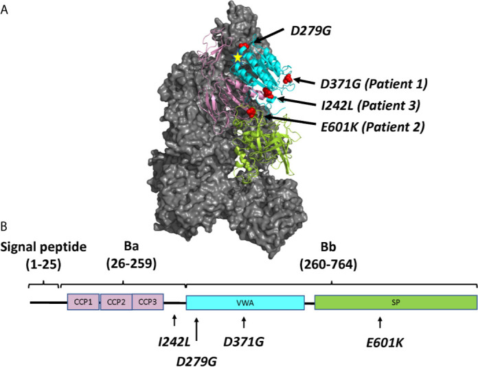
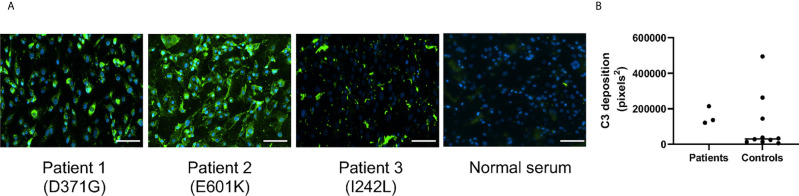
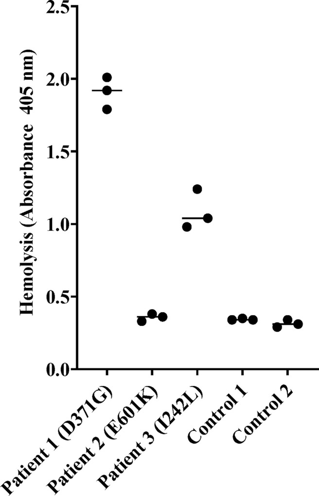
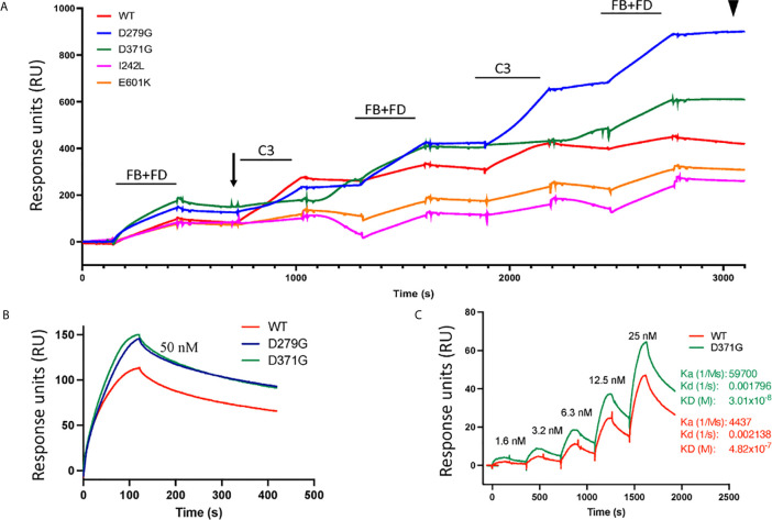
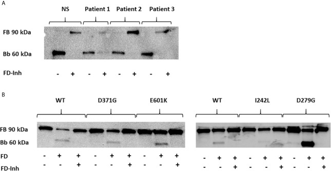
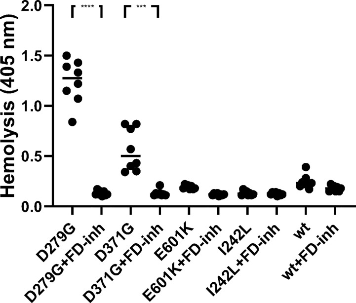
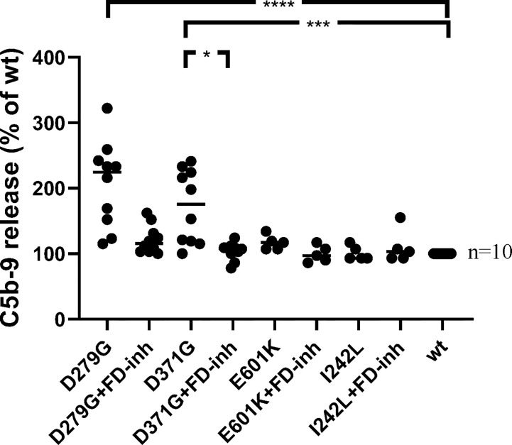
Complement factor B (FB) mutant variants are associated with excessive complement activation in kidney diseases such as atypical hemolytic uremic syndrome (aHUS), C3 glomerulopathy and membranoproliferative glomerulonephritis (MPGN). Patients with aHUS are currently treated with eculizumab while there is no specific treatment for other complement-mediated renal diseases. In this study the phenotype of three FB missense variants, detected in patients with aHUS (D371G and E601K) and MPGN (I242L), was investigated. Patient sera with the D371G and I242L mutations induced hemolysis of sheep erythrocytes. Mutagenesis was performed to study the effect of factor D (FD) inhibition on C3 convertase-induced FB cleavage, complement-mediated hemolysis, and the release of soluble C5b-9 from glomerular endothelial cells. The FD inhibitor danicopan abrogated C3 convertase-associated FB cleavage to the Bb fragment in patient serum, and of the FB constructs, D371G, E601K, I242L, the gain-of-function mutation D279G, and the wild-type construct, in FB-depleted serum. Furthermore, the FD-inhibitor blocked hemolysis induced by the D371G and D279G gain-of-function mutants. In FB-depleted serum the D371G and D279G mutants induced release of C5b-9 from glomerular endothelial cells that was reduced by the FD-inhibitor. These results suggest that FD inhibition can effectively block complement overactivation induced by FB gain-of-function mutations.

Keywords: C3 glomerulopathy; atypical hemolytic uremic syndrome; complement; danicopan; factor B; factor D.


Conflict of interest statement

The authors declare that the research was conducted in the absence of any commercial or financial relationships that could be construed as a potential conflict of interest.

Figures

Figure 1
Figure 1
The molecular structure of factor B and the location of mutations described in this study. (A) Location of the four mutated residues (D371G, E601K, I242L and D279G), visualized on the structure of the C3 pro-convertase C3bB depicted in grey (PDB ID 2XWJ) (18) using PyMol. The Metal Ion Dependent Adhesion Site (MIDAS) on C3b is depicted by a star. The colors of the domains correspond to the domains depicted in (B). (B) The linear structure of factor B divided into the signal peptide/leader, Ba and Bb fragments with a linker sequence in between. The amino acid numbers are given in parentheses. The protein is composed of a complement control protein (CCP) domain with three CCPs, followed by the linker, the von Willebrand factor type A domain (VWA) and the serine protease (SP) domain. The location of mutations studied herein is depicted.
Figure 2
Figure 2
C3 deposition on glomerular endothelial cells in the presence of serum from patients and controls. Sera from Patients 1-3 and controls (n = 10) were incubated with glomerular endothelial cells for 2 h and C3 detected by immunofluorescence. (A) Serum from Patients 1-3 induced excessive C3 deposition on the cells. The scale bar represents 100 µm. (B) Quantification of C3 deposition fluorescence showing that even 3/10 sera from apparently healthy adult controls exhibited C3 deposition. The bar represents the median.
Figure 3
Figure 3
Hemolysis of sheep erythrocytes in the presence of patient and normal serum. Serum from Patients 1 (factor B mutation D371G) and 3 (I242L) induced hemolysis of sheep erythrocytes whereas serum from Patient 2 and from the two normal controls did not. The results of three separate experiments are shown. The bar depicts the median.
Figure 4
Figure 4
Binding of factor B variants to C3b and formation of the C3 convertase. (A) Purified C3b was coupled to a CM5 sensor chip. Factor B (FB) variants and factor D (FD) were injected over the surface and binding curves visualized. The FB D371G mutant exhibited the strongest binding to the C3b-coated surface (see arrow) followed by D279G, I242L, the wild-type (WT) and E601K. This was followed by serial injections of C3 alternating with FB+FD to form the C3 convertase on the chip. The strongest C3 convertase generation was demonstrated for the D279G mutant (see arrowhead), followed by D371G, the wild-type, I242L and E601K. Baseline values were adjusted at t = 0 in each cycle for comparison. (B) Binding between C3b and FB alone was assessed using the wild-type construct, D371G and D279G showing that both mutant constructs, at 50 nM, exhibited stronger binding than the wild-type construct. (C) The coefficient of dissociation was evaluated using a range of FB concentrations comparing construct D371G to the wild-type.
Figure 5
Figure 5
The effect of factor D inhibition on C3 convertase formation using human serum or factor B mutants. An immunoblot assay was used to detect the Bb fragment of the alternative pathway C3bBb. (A) C3bBb(Mg2+) complexes were formed by incubating C3b-coated wells with normal serum (NS) or patient serum (Patients 1-3). The C3 convertase formed in the presence of all sera effectively cleaved factor B to the Bb component and this reaction was inhibited by the factor D inhibitor. The factor D inhibitor only partially blocked the C3 convertase in the presence of serum from Patient 1(D371G mutation) as a weak Bb band was still visible. (B) The same assay was performed with the wild-type (WT) and mutant factor B constructs (D371G, E601K, I242L and D279G) showing cleavage to the Bb fragments and effective inhibition by the factor D inhibitor. FB, factor B; Bb, the Bb fragment of factor B; FD, factor D; FD-inh, factor D inhibitor.
Figure 6
Figure 6
The effect of factor D inhibition on hemolysis of rabbit erythrocytes. Factor B constructs were incubated in factor B-depleted serum with rabbit erythrocytes. The mutant variants D279G (positive control) and D371G (corresponding to Patient 1) induced hemolysis. The other mutant constructs (E601K, I242L) and wild-type (wt) did not induce hemolysis. The factor D inhibitor (FD-inh) inhibited hemolysis induced by factor B mutants D279G and D371G. Eight separate experiments are shown. ***P < 0.001, ****P < 0.0001.
Figure 7
Figure 7
The effect of factor D inhibition on C5b-9 release from glomerular endothelial cells. Factor B mutants D279G (positive control), D371G, E601K, I242L and the wild-type (wt) construct were combined with factor B-depleted serum and incubated with glomerular endothelial cells. The release of C5b-9 was detected in cell supernatants in the presence or absence of the factor D inhibitor. The mutant construct D371G induced the release of C5b-9 which was decreased by the factor D inhibitor. A tendency to decrease was noted when the mutant construct D279G was incubated with the factor D inhibitor but this did not achieve statistical significance (multivariate analysis). FD-inh, factor D inhibitor; *P < 0.05, ***P < 0.001, ****P < 0.0001.

References

    1. Walport MJ. Complement. First of Two Parts. N Engl J Med (2001) 344:1058–66. 10.1056/NEJM200104053441406 - DOI - PubMed
    1. Slade C, Bosco J, Unglik G, Bleasel K, Nagel M, Winship I. Deficiency in Complement Factor B. N Engl J Med (2013) 369:1667–9. 10.1056/NEJMc1306326 - DOI - PubMed
    1. Karpman D, Loos S, Tati R, Arvidsson I. Haemolytic Uraemic Syndrome. J Intern Med (2017) 281:123–48. 10.1111/joim.12546 - DOI - PubMed
    1. Smith RJH, Appel GB, Blom AM, Cook HT, D’Agati VD, Fakhouri F, et al. . C3 Glomerulopathy - Understanding a Rare Complement-Driven Renal Disease. Nat Rev Nephrol (2019) 15:129–43. 10.1038/s41581-018-0107-2 - DOI - PMC - PubMed
    1. Goodship TH, Cook HT, Fakhouri F, Fervenza FC, Fremeaux-Bacchi V, Kavanagh D, et al. . Atypical Hemolytic Uremic Syndrome and C3 Glomerulopathy: Conclusions From a “Kidney Disease: Improving Global Outcomes” (Kdigo) Controversies Conference. Kidney Int (2017) 91:539–51. 10.1016/j.kint.2016.10.005 - DOI - PubMed

Publication types

MeSH terms