

Structure of Blood Coagulation Factor VIII in Complex With an Anti-C2 Domain Non-Classical, Pathogenic Antibody Inhibitor
- PMID: 34177966
- PMCID: PMC8223065
- DOI: 10.3389/fimmu.2021.697602
Structure of Blood Coagulation Factor VIII in Complex With an Anti-C2 Domain Non-Classical, Pathogenic Antibody Inhibitor
Abstract
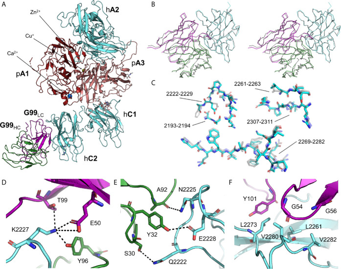
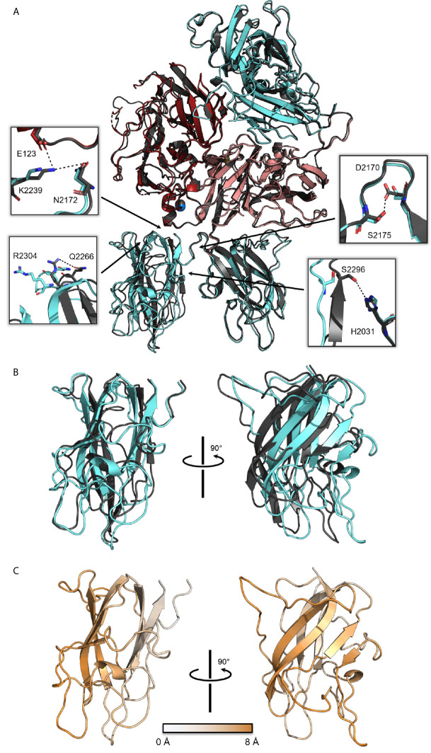
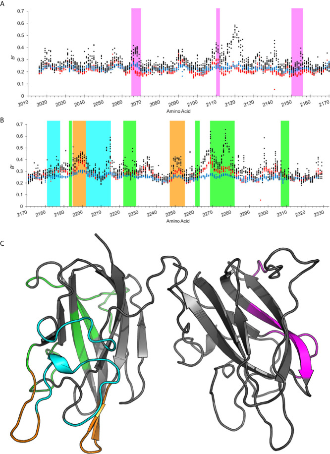
Factor VIII (fVIII) is a procoagulant protein that binds to activated factor IX (fIXa) on platelet surfaces to form the intrinsic tenase complex. Due to the high immunogenicity of fVIII, generation of antibody inhibitors is a common occurrence in patients during hemophilia A treatment and spontaneously occurs in acquired hemophilia A patients. Non-classical antibody inhibitors, which block fVIII activation by thrombin and formation of the tenase complex, are the most common anti-C2 domain pathogenic inhibitors in hemophilia A murine models and have been identified in patient plasmas. In this study, we report on the X-ray crystal structure of a B domain-deleted bioengineered fVIII bound to the non-classical antibody inhibitor, G99. While binding to G99 does not disrupt the overall domain architecture of fVIII, the C2 domain undergoes an ~8 Å translocation that is concomitant with breaking multiple domain-domain interactions. Analysis of normalized B-factor values revealed several solvent-exposed loops in the C1 and C2 domains which experience a decrease in thermal motion in the presence of inhibitory antibodies. These results enhance our understanding on the structural nature of binding non-classical inhibitors and provide a structural dynamics-based rationale for cooperativity between anti-C1 and anti-C2 domain inhibitors.
Keywords: antibody binding; antibody inhibitors; blood coagulation; factor VIII; x-ray crystallography.
Copyright © 2021 Ronayne, Peters, Gish, Wilson, Spencer, Doering, Lollar, Spiegel and Childers.
Conflict of interest statement
PL is inventor on a patent application describing ET3i and is an inventor on patents owned by Emory University claiming compositions of matter that include modified fVIII proteins with reduced reactivity with anti-fVIII antibodies. CD, PL and HS are cofounders of Expression Therapeutics and own equity in the company. Expression Therapeutics owns the intellectual property associated with ET3i. The terms of this arrangement have been reviewed and approved by Emory University in accordance with its conflict of interest policies. The remaining authors declare that the research was conducted in the absence of any commercial or financial relationships that could be construed as a potential conflict of interest.
Figures
Similar articles
-
Structure of blood coagulation factor VIII in complex with an anti-C1 domain pathogenic antibody inhibitor.Blood. 2021 May 27;137(21):2981-2986. doi: 10.1182/blood.2020008940. Blood. 2021. PMID: 33529335 Free PMC article.
-
High-affinity, noninhibitory pathogenic C1 domain antibodies are present in patients with hemophilia A and inhibitors.Blood. 2016 Oct 20;128(16):2055-2067. doi: 10.1182/blood-2016-02-701805. Epub 2016 Jul 5. Blood. 2016. PMID: 27381905 Free PMC article.
-
The 1.7 Å X-ray crystal structure of the porcine factor VIII C2 domain and binding analysis to anti-human C2 domain antibodies and phospholipid surfaces.PLoS One. 2015 Mar 16;10(3):e0122447. doi: 10.1371/journal.pone.0122447. eCollection 2015. PLoS One. 2015. PMID: 25775247 Free PMC article.
-
Epitope specificity and inactivation mechanisms of factor VIII inhibitor antibodies.Vox Sang. 1999;77 Suppl 1:17-20. doi: 10.1159/000056708. Vox Sang. 1999. PMID: 10529680 Review.
-
Structural insights into blood coagulation factor VIII: Procoagulant complexes, membrane binding, and antibody inhibition.J Thromb Haemost. 2022 Sep;20(9):1957-1970. doi: 10.1111/jth.15793. Epub 2022 Jul 11. J Thromb Haemost. 2022. PMID: 35722946 Review.
Cited by
-
Genetics and Epigenetics in Acquired Hemophilia A: From Bench to Bedside.Curr Issues Mol Biol. 2024 May 23;46(6):5147-5160. doi: 10.3390/cimb46060309. Curr Issues Mol Biol. 2024. PMID: 38920981 Free PMC article. Review.
-
SAXS analysis of the intrinsic tenase complex bound to a lipid nanodisc highlights intermolecular contacts between factors VIIIa/IXa.Blood Adv. 2022 Jun 14;6(11):3240-3254. doi: 10.1182/bloodadvances.2021005874. Blood Adv. 2022. PMID: 35255502 Free PMC article.
-
Biophysical characterization of blood coagulation factor VIII binding to lipid nanodiscs that mimic activated platelet surfaces.J Thromb Haemost. 2025 Feb;23(2):513-524. doi: 10.1016/j.jtha.2024.11.003. Epub 2024 Nov 15. J Thromb Haemost. 2025. PMID: 39549835
-
Structural basis for inhibition of coagulation factor VIII reveals a shared antigenic hotspot on the C1 domain.J Thromb Haemost. 2024 Sep;22(9):2449-2459. doi: 10.1016/j.jtha.2024.05.024. Epub 2024 Jun 5. J Thromb Haemost. 2024. PMID: 38849084 Free PMC article.
-
An intronic RNA element modulates Factor VIII exon-16 splicing.Nucleic Acids Res. 2024 Jan 11;52(1):300-315. doi: 10.1093/nar/gkad1034. Nucleic Acids Res. 2024. PMID: 37962303 Free PMC article.
References
Publication types
MeSH terms
Substances
Supplementary concepts
Grants and funding
LinkOut - more resources
Full Text Sources
Molecular Biology Databases
Miscellaneous