

ADSC Exosomes Mediate lncRNA-MIAT Alleviation of Endometrial Fibrosis by Regulating miR-150-5p
- PMID: 34178037
- PMCID: PMC8220143
- DOI: 10.3389/fgene.2021.679643
ADSC Exosomes Mediate lncRNA-MIAT Alleviation of Endometrial Fibrosis by Regulating miR-150-5p
Abstract
Background: Secondary infertility remains a major complication of endometrial fibrosis in women. The use of exosomes from adipose-derived mesenchymal stem cells (ADSCs) has shown promising results for the treatment of endometrial fibrosis. However, the mechanisms of action of ADSC-exosome (ADSC-Exo) therapy remain unclear.
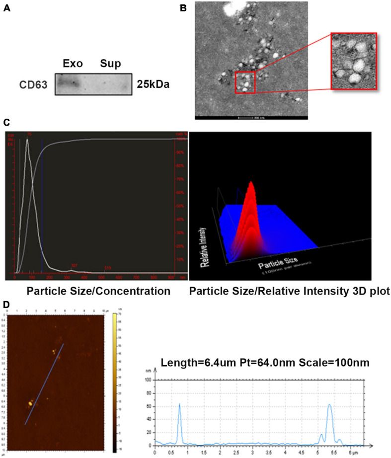
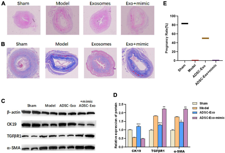
Materials and methods: An endometrial fibrosis model was established in mice treated with alcohol and endometrial epithelial cells (ESCs) treated with TGF-β1. ADSCs were isolated from Sprague Dawley (SD) rats, and exosomes were isolated from ADSCs using ExoQuick reagent. Exosomes were identified by transmission electron microscopy (TEM), NanoSight, and Western blot analysis. The expression level of lncRNA-MIAT was detected by qPCR analysis. Western blot analysis was carried out to determine the protein levels of fibrosis markers (TGFβR1, α-SMA, and CK19). A dual-luciferase reporter gene assay was used to verify the relationship between target genes. The endometrial tissues of the endometrial fibrosis model were stained with HE and Masson's trichrome.
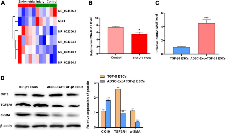
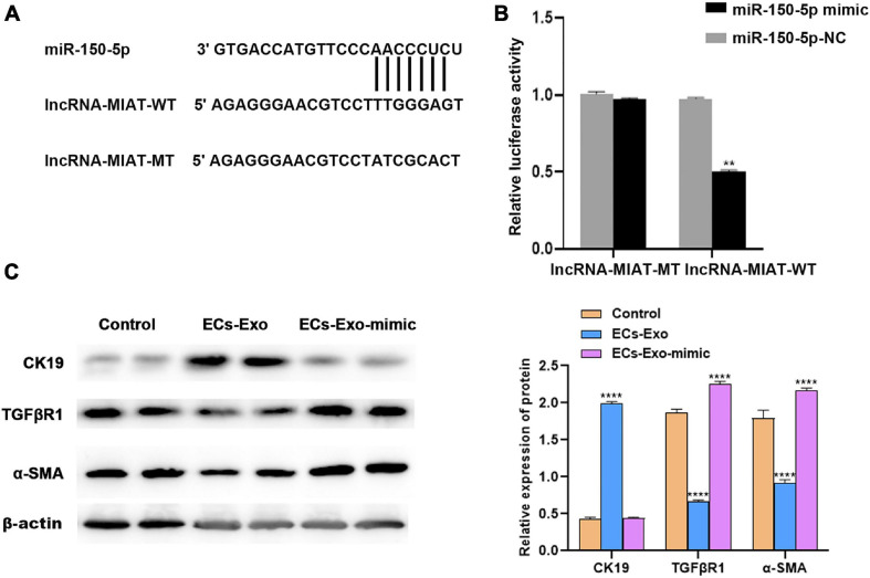
Results: ADSCs and ADSC-Exos were successfully isolated, and the expression level of lncRNA-MIAT was significantly down-regulated in endometrial tissue and the TGF-β1-induced ESC injury model, whereas ADSC-Exos increased the expression of lncRNA-MIAT in the TGF-β1-induced ESC model. Functionally, ADSC-Exo treatment repressed endometrial fibrosis in vivo and in vitro by decreasing the expression of hepatic fibrosis markers (α-SMA and TGFβR1) and increasing the expression of CK19. Moreover, miR-150-5p expression was repressed by lncRNA-MIAT in the TGF-β1-induced ESC injury model. The miR-150-5p mimic promoted TGF-β1-induced ESC fibrosis.
Conclusion: ADSC-Exos mediate lncRNA-MIAT alleviation of endometrial fibrosis by regulating miR-150-5p, which suggests that lncRNA-MIAT from ADSC-Exos may be a viable treatment for endometrial fibrosis.
Keywords: ADSC; ADSC-exosomes; LncRNA-MIAT; endometrial fibrosis; miR-150-5p.
Copyright © 2021 Shao, Qin, Wan, Cheng, Wang, Ai, Cheng and Tong.
Conflict of interest statement
The authors declare that the research was conducted in the absence of any commercial or financial relationships that could be construed as a potential conflict of interest.
Figures
Similar articles
-
Exosomes from adipose-derived mesenchymal stem cells improve liver fibrosis by regulating the miR-20a-5p/TGFBR2 axis to affect the p38 MAPK/NF-κB pathway.Cytokine. 2023 Dec;172:156386. doi: 10.1016/j.cyto.2023.156386. Epub 2023 Oct 16. Cytokine. 2023. PMID: 37852157
-
Exosomes from adipose-derived mesenchymal stem cell improve diabetic wound healing and inhibit fibrosis via miR-128-1-5p/TGF-β1/Smad axis.Mol Cell Endocrinol. 2024 Jul 1;588:112213. doi: 10.1016/j.mce.2024.112213. Epub 2024 Mar 29. Mol Cell Endocrinol. 2024. PMID: 38556162
-
Exosomes Derived from Human Adipose Mesenchymal Stem Cells Inhibits Fibrosis and Treats Oral Submucous Fibrosis via the miR-181a-5p/Smad2 Axis.Tissue Eng Regen Med. 2024 Jan;21(1):123-135. doi: 10.1007/s13770-023-00579-0. Epub 2023 Sep 27. Tissue Eng Regen Med. 2024. PMID: 37755664 Free PMC article.
-
Adipose-Derived Stem Cell Exosomes as a Novel Anti-Inflammatory Agent and the Current Therapeutic Targets for Rheumatoid Arthritis.Biomedicines. 2022 Jul 18;10(7):1725. doi: 10.3390/biomedicines10071725. Biomedicines. 2022. PMID: 35885030 Free PMC article. Review.
-
Therapeutic potential of exosomes from adipose-derived stem cells in chronic wound healing.Front Surg. 2022 Sep 30;9:1030288. doi: 10.3389/fsurg.2022.1030288. eCollection 2022. Front Surg. 2022. PMID: 36248361 Free PMC article. Review.
Cited by
-
MSC-derived exosomes improve endometrial fibrosis via the lncRNA IGF2R/ miR-143-5p/AQP8 axis.Stem Cell Res Ther. 2025 Aug 27;16(1):464. doi: 10.1186/s13287-025-04611-z. Stem Cell Res Ther. 2025. PMID: 40866943 Free PMC article.
-
RNA sequencing of exosomes secreted by fibroblast and Schwann cells elucidates mechanisms underlying peripheral nerve regeneration.Neural Regen Res. 2024 Aug 1;19(8):1812-1821. doi: 10.4103/1673-5374.387980. Epub 2023 Nov 8. Neural Regen Res. 2024. PMID: 38103248 Free PMC article.
-
Effects of ADSC-Derived Exosome LRRC75A-AS1 on Anti-inflammatory Function After SCI.Appl Biochem Biotechnol. 2024 Sep;196(9):5920-5935. doi: 10.1007/s12010-023-04836-9. Epub 2024 Jan 2. Appl Biochem Biotechnol. 2024. PMID: 38165592
-
RGD hydrogel-loaded ADSC extracellular vesicles mitigate uranium-induced renal injury via TLR4/NF-κB pathway inhibition.J Nanobiotechnology. 2025 Feb 17;23(1):114. doi: 10.1186/s12951-025-03176-6. J Nanobiotechnology. 2025. PMID: 39962465 Free PMC article.
-
Extracellular Vesicles Derived from Mesenchymal Stem Cells as Cell-Free Therapy for Intrauterine Adhesion.Int J Stem Cells. 2023 Aug 30;16(3):260-268. doi: 10.15283/ijsc21177. Epub 2023 Jun 30. Int J Stem Cells. 2023. PMID: 37385632 Free PMC article. Review.
References
LinkOut - more resources
Full Text Sources