A Case of Coronary Cameral Fistula: When and How to Intervene?
- PMID: 34178089
- PMCID: PMC8217193
- DOI: 10.18502/jthc.v15i4.5946
A Case of Coronary Cameral Fistula: When and How to Intervene?
Abstract
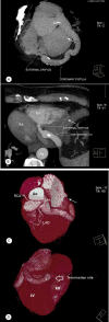
Coronary artery fistulas constitute a rare anomaly defined as an abnormal communication between a coronary artery and a great vessel or any cardiac chamber. The majority of these fistulas arise from the right coronary artery and the left anterior descending coronary artery; the circumflex coronary artery is rarely involved. We present an unusual case of a coronary artery fistula in a middle-aged woman who presented with symptoms of heart failure and abnormal auscultation. Echocardiography and conventional and computed tomography angiography showed that the coronary fistula originated from the left circumflex coronary artery and drained majorly into the right ventricle. Given the complex anatomy of the fistula, we managed it surgically rather than percutaneously. There were no complications early after surgery and at 1 year's follow-up.
Keywords: Computed tomography angiography; Fistula; Heart Defects* Congenital.
Copyright © 2020 Tehran University of Medical Sciences.
Figures


References
-
- Pompa JJ, Kinlay S, Bhatt DL. Coronary arteriography and intracoronary imaging. In: Bonow RO, Mann DL, Zipes DP, Libby P, Braunwald E, eds , editors. Braunwald’s Heart Disease: A Textbook of Cardiovascular Medicine. 9th ed. . Philadelphia: Saunders; 2015. pp. 392–424.
-
- Gowda RM, Vasavada BC, Khan IA. Coronary artery fistulas: clinical and therapeutic considerations. Int J Cardiol. 2006;107:7–10. - PubMed
-
- Zenooz NA, Habibi R, Mammen L, Finn JP, Gilkeson RC. Coronary artery fistulas: CT findings. Radiographics. 2009;29:781–789. - PubMed
-
- Lim JJ, Jung JI, Lee BY, Lee HG. Prevalence and types of coronary artery fistulas detected with coronary CT angiography. AJR Am J Roentgenol. 2014;203:W237–243. - PubMed
-
- Greenberg MA, Fish BG, Spindola-Franco H. Congenital anomalies of coronary artery: classification and significance. Radiol Clin North Am. 1989;27:1127–1146. - PubMed
LinkOut - more resources
Full Text Sources
