Case Reports
. 2021 Jun 12;16(8):2192-2201.
doi: 10.1016/j.radcr.2021.05.045. eCollection 2021 Aug.

Sarcoidosis-lymphoma syndrome with portal hypertension: diagnostic clues and approach

Affiliations
Case Reports

Sarcoidosis-lymphoma syndrome with portal hypertension: diagnostic clues and approach

Fumio Chikamori et al. Radiol Case Rep. .

Abstract

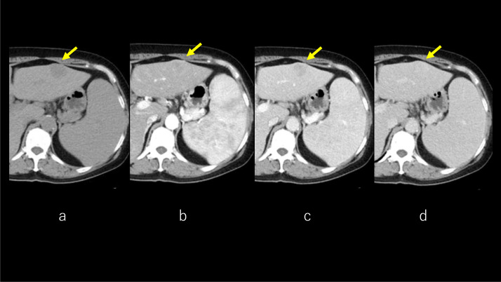
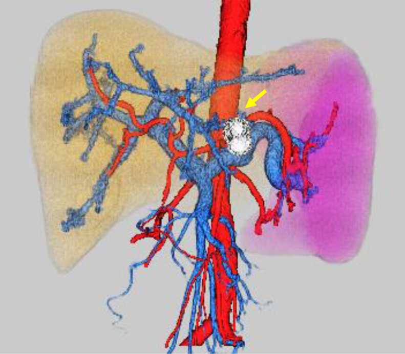
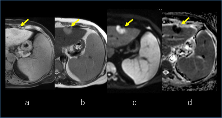
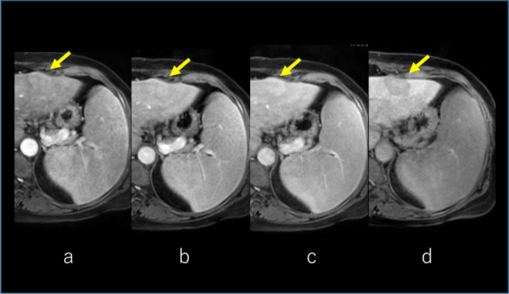
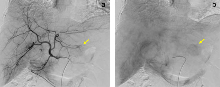
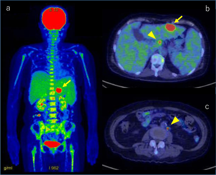
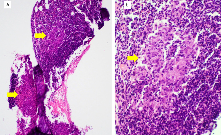
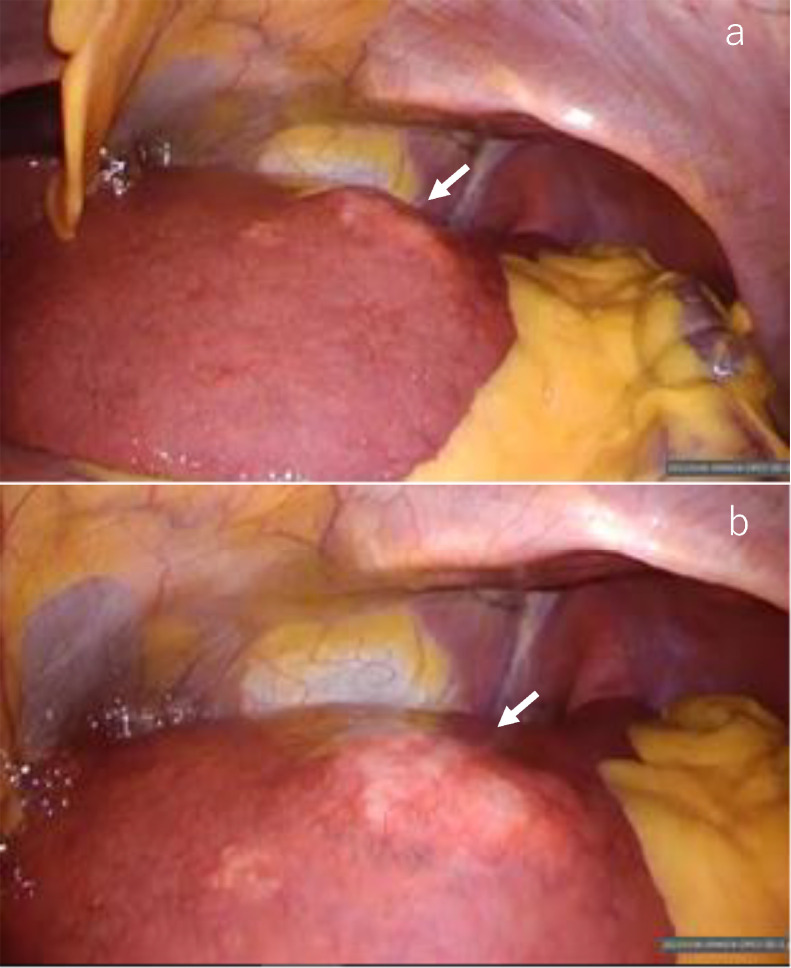
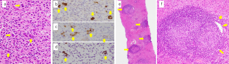
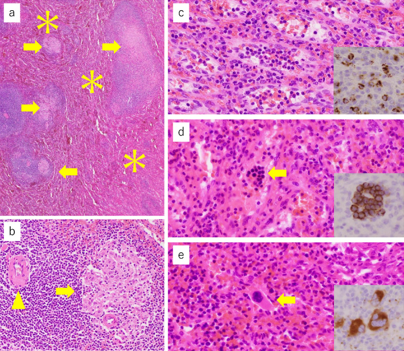
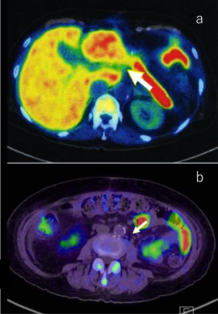
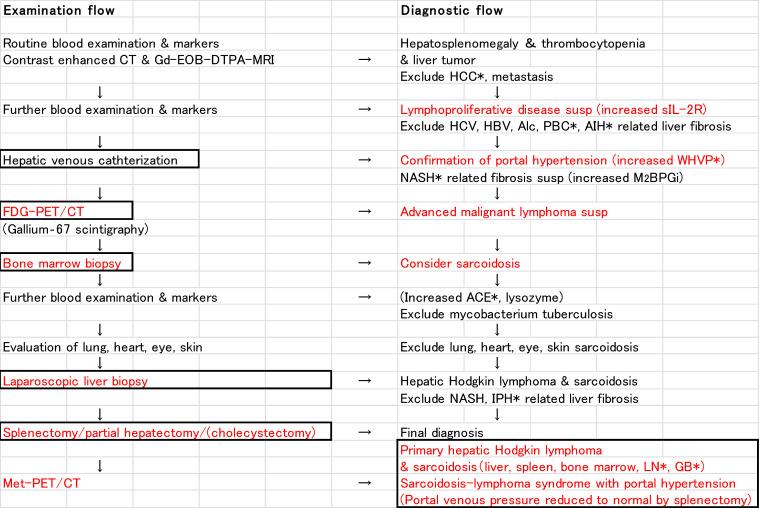
Sarcoidosis-lymphoma syndrome associated with portal hypertension is very rare. A 68-year-old female presented with a 5 kg weight loss in 6 months. Soluble interleukin-2 receptor activity was increased and total platelet count was decreased. Contrast-enhanced computed tomography showed the presence of hepatosplenomegaly and a 3 cm-sized tumor in segment 3 of the liver. The hepatic venous catheterization showed mild portal hypertension. On fluorodeoxyglucose-positron emission tomography/computed tomography, progressive malignant lymphoma was suspected. However, bone marrow biopsy showed multiple noncaseating granulomas. A laparoscopic liver biopsy revealed that the liver tumor had features of Hodgkin lymphoma. There were multiple noncaseating epithelioid granulomas in the portal tracts of the liver. Splenectomy for splenomegaly and partial hepatectomy for the liver tumor were performed. Pathological examination of the resected specimens revealed multiple noncaseating epithelioid granulomas in the liver and spleen. Histopathology of the liver tumor confirmed classic Hodgkin lymphoma with mixed cellularity. We conclude that hepatic venous catheterization, positron emission tomography/computed tomography, and pathological examinations of bone marrow, liver, and spleen are crucial for the diagnosis of sarcoidosis-lymphoma syndrome associated with portal hypertension.

Keywords: Hepatic sarcoidosis; Laparoscopic liver biopsy; PET/CT; Portal hypertension; Primary hepatic Hodgkin lymphoma; Sarcoidosis-lymphoma syndrome.


Figures

Fig 1
Fig. 1
CT. Abdominal plain CT shows solitary, hypoattenuating tumor (arrow) in the left lobe (A). The arterial phase of dynamic CT shows tumor enhancement with central part sparing (B). The tumor is demonstrated clearly in the portal phase (C). The equilibrium phase of dynamic CT shows the lesion with persistent enhancement (D). CT, computed tomography.
Fig 2
Fig. 2
3D-CT reconstruction image. 3D-CT reconstruction image shows hepatosplenomegaly and increased splenic vein diameter. The spleen volume is 1011 mL and the liver volume is 2461 mL, respectively. Spleen/liver volume ratio is 0.41. No development of portal collaterals is observed. The tumor (arrow) is located in segment 3. CT, computed tomography.
Fig 3
Fig. 3
MRI of the liver. The tumor was visible in the left lobe which presents hypointense in T1-weighted image (arrow) (A) and slightly hyperintense in T2-weighted image (arrow) (B). The lesion was presenting with a high signal in the diffusion-weighted image (DWI) (arrow) (C) and a low signal in the apparent diffusion coefficient (ADC) map (arrow) (D). MRI, magnetic resonance imaging.
Fig 4
Fig. 4
Gd-EOB-DTPA-enhanced MRI. Arterial, portal, and transitional phases of the dynamic scan show enhancement of the tumor with central part sparing (arrow) (A, B, C). The hepatobiliary phase shows a faint uptake of contrast in the central part of the tumor (arrow) (D). MRI, magnetic resonance imaging.
Fig 5
Fig. 5
Hepatic arteriography. Early (A) and late (B) phases of hepatic arteriography show fine tumor staining (arrow) in segment 3.
Fig 6
Fig. 6
Whole-body scan of FDG-PET/CT. Coronal fusion image shows intensive uptake in the hepatic tumor (arrow), with an SUV max of 21.0 in segment 3 (A). There is diffuse hyperaccumulation in the bone marrow of the spine, pelvic bones, ribs, and upper and lower limbs (SUV max, 5.6) (A). Axial fusion image shows lymph nodes (arrowhead) around the hepatic artery (B) and around the aorta (C) with intensive uptake (SUV max, 21.9). FDG-PET/CT, fluorodeoxyglucose-positron emission tomography/computed tomography.
Fig 7
Fig. 7
Histopathological findings of the biopsied bone marrow. The bone marrow shows multiple noncaseating epithelioid granulomas (A, lower magnification, ×40; B, higher magnification, ×400; granulomas indicated by arrows).
Fig 8
Fig. 8
Laparoscopic view. Laparoscopic distal (A) and close-up views (B) show a slightly protruding white nodule (arrow).
Fig 9
Fig. 9
Histopathological findings of the liver biopsy. High magnification view shows scattered localization of Reed-Sternberg cells (A, ×400, arrows) which are immunohistochemically positive for PAX5 (B, ×400, arrows) and CD30 (C, ×400, arrows) and positive for Epstein-Barr virus-encoded small RNA 1 by in situ hybridization (D, ×400, arrows). The background hepatic parenchyma shows noncaseating epithelioid granulomas (arrows) developed mainly in the portal triads (E, ×40). The higher magnification view of the portal triad (F, ×200) including an epithelioid granuloma shows that the granuloma largely occupying the triad pushes bile ducts (arrows) and portal venule (arrowhead) to the periphery. The portal venule appears to be compressed by inflammation.
Fig 10
Fig. 10
Histopathological findings of the resected spleen. The spleen pathologically shows that multiple noncaseating epithelioid granulomas (A, B, arrows) are mainly present in the white pulps (B, a splenic central artery is indicated by an arrowhead) and red pulps (asterisks) are extended (A, ×40). Red pulps demonstrate extramedullary hematopoiesis because, in red pulps, there are neutrophils (C, ×400) and myeloperoxidase-immunoreactive myeloid cells (C, inset, ×400), erythroblastic islands (D, ×400) that are immunohistochemically positive for CD71 (D, inset, ×400), and megakaryocytes (E, ×400) that are immunohistochemically positive for CD61 (E, inset, ×400).
Fig 11
Fig. 11
Axial fusion image of MET-PET/CT after the surgery. Lymph nodes around the hepatic artery (A) and around the aorta (B), which showed high accumulation on FDP-PET/CT, show low accumulation on MET-PET/CT (SUV max, 3.7 and 1.3, respectively). The findings are consistent with sarcoidosis. MET-PET/CT, methionine-positron emission tomography/computed tomography.
Fig 12
Fig. 12
Examination and diagnostic flow of the case. HCC, hepatocellular carcinoma; PBC, primary biliary cirrhosis; AIH, autoimmune hepatitis; WHVP, wedged hepatic venous pressure; ACE, angiotensin-converting enzyme; IPH, idiopathic portal hypertension; LN, lymphnodes; GB, gallbladder.

Similar articles

Cited by

References

    1. Brincker H. The sarcoidosis-lymphoma syndrome. Br J Cancer. 1986;54(3):467–473. doi: 10.1038/bjc.1986.199. - DOI - PMC - PubMed
    1. Papanikolaou IC, Sharma OP. The relationship between sarcoidosis and lymphoma. Eur Respir J. 2010;36(5):1207–1219. doi: 10.1183/09031936.00043010. - DOI - PubMed
    1. Oskuei A, Hicks L, Ghaffar H, Hoffstein V. Sarcoidosis-lymphoma syndrome: a diagnostic dilemma. BMJ Case Rep. 2017;2017 doi: 10.1136/bcr-2017-220065. - DOI - PMC - PubMed
    1. Yamanaka T, Kanai H, Aihara N, Ohno T, Mase M. A case of sarcoidosis-lymphoma syndrome: importance of brain biopsy. NMC Case Report J. 2019;6(2):61–64. doi: 10.2176/nmccrj.cr.2018-0223. - DOI - PMC - PubMed
    1. Kis A, Eszes N, Tamasi L, Losonczy G, Csekeo A, Csomor J. Sarcoidosis lymphoma syndrome—the value of PET-CT in the diagnosis. World J Sur Oncol. 2013;11:235. doi: 10.1186/1477-7819-11-235. - DOI - PMC - PubMed

Publication types

LinkOut - more resources