

Covid-19 imaging: A narrative review
- PMID: 34178312
- PMCID: PMC8214462
- DOI: 10.1016/j.amsu.2021.102489
Covid-19 imaging: A narrative review
Abstract
Background: The 2019 novel coronavirus disease (COVID-19) imaging data is dispersed in numerous publications. A cohesive literature review is to be assembled.
Objective: To summarize the existing literature on Covid-19 pneumonia imaging including precautionary measures for radiology departments, Chest CT's role in diagnosis and management, imaging findings of Covid-19 patients including children and pregnant women, artificial intelligence applications and practical recommendations.
Methods: A systematic literature search of PubMed/med line electronic databases.
Results: The radiology department's staff is on the front line of the novel coronavirus outbreak. Strict adherence to precautionary measures is the main defense against infection's spread. Although nucleic acid testing is Covid-19's pneumonia diagnosis gold standard; kits shortage and low sensitivity led to the implementation of the highly sensitive chest computed tomography amidst initial diagnostic tools. Initial Covid-19 CT features comprise bilateral, peripheral or posterior, multilobar ground-glass opacities, predominantly in the lower lobes. Consolidations superimposed on ground-glass opacifications are found in few cases, preponderantly in the elderly. In later disease stages, GGO transformation into multifocal consolidations, thickened interlobular and intralobular lines, crazy paving, traction bronchiectasis, pleural thickening, and subpleural bands are reported. Standardized CT reporting is recommended to guide radiologists. While lung ultrasound, pulmonary MRI, and PET CT are not Covid-19 pneumonia's first-line investigative diagnostic modalities, their characteristic findings and clinical value are outlined. Artificial intelligence's role in strengthening available imaging tools is discussed.
Conclusion: This review offers an exhaustive analysis of the current literature on imaging role and findings in COVID-19 pneumonia.
Keywords: COVID-19; Chest; Computed tomography (CT); Imaging; Novel coronavirus; Sars-Cov2.
© 2021 The Authors.
Conflict of interest statement
Authors have no conflicts of interest. No funding was received.
Figures


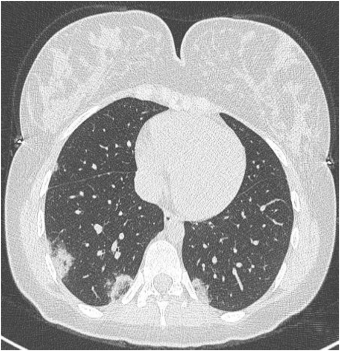
(a) A left upper lobe unifocal rounded ground glass opacity;
(b) Patchy peripheral ground glass opacity with vascular dilatation (Black circle);
(c) Multifocal, bilateral subpleural ground glass opacities with traction bronchiectasis (Black circle);
(d) Extensive bilateral ground glass opacities associated with thickened interlobular and intralobular septa (Crazy paving) alongside with peripheral consolidations;
(e) Subpleural band in advanced-phase disease (Black arrow).

(a,b) Early stage: Ground glass opacities involving the lower lobes with partial crazy paving;
(c) Progressive stage: Ground glass opacities extension and increased crazy paving;
(d,e) Peak stage: Consolidative opacities, sub-pleural lines (Black arrow) and bronchiectasis.


(a,b) Acute eosinophilic pneumonia associating interlobular septal thickening, lower lobes air space consolidations and pleural effusions (*).
(c,d) Extensive, multifocal, bilateral consolidation in a case of diffuse alveolar hemorrhage.

Similar articles
-
Coronavirus Disease 2019 (COVID-19): A Systematic Review of Imaging Findings in 919 Patients.AJR Am J Roentgenol. 2020 Jul;215(1):87-93. doi: 10.2214/AJR.20.23034. Epub 2020 Mar 14. AJR Am J Roentgenol. 2020. PMID: 32174129
-
COVID-19 pneumonia manifestations at the admission on chest ultrasound, radiographs, and CT: single-center study and comprehensive radiologic literature review.Eur J Radiol Open. 2020;7:100231. doi: 10.1016/j.ejro.2020.100231. Epub 2020 Apr 4. Eur J Radiol Open. 2020. PMID: 32289051 Free PMC article.
-
Rapid onset of bronchiectasis in COVID-19 Pneumonia: two cases studied with CT.Radiol Case Rep. 2020 Nov;15(11):2098-2103. doi: 10.1016/j.radcr.2020.08.008. Epub 2020 Aug 5. Radiol Case Rep. 2020. PMID: 32837670 Free PMC article.
-
COVID-19 integrated imaging: our experience and literature review.Pol J Radiol. 2021 Feb 1;86:e78-e86. doi: 10.5114/pjr.2021.103861. eCollection 2021. Pol J Radiol. 2021. PMID: 33758632 Free PMC article.
-
Computed Tomography (CT) Imaging Features of Patients with COVID-19: Systematic Review and Meta-Analysis.Radiol Res Pract. 2020 Jul 23;2020:1023506. doi: 10.1155/2020/1023506. eCollection 2020. Radiol Res Pract. 2020. PMID: 32733706 Free PMC article. Review.
Cited by
-
CovidConvLSTM: A fuzzy ensemble model for COVID-19 detection from chest X-rays.Expert Syst Appl. 2022 Nov 15;206:117812. doi: 10.1016/j.eswa.2022.117812. Epub 2022 Jun 16. Expert Syst Appl. 2022. PMID: 35754941 Free PMC article.
-
Retrospective Cohort Study Comparing the Clinical Profile and Outcomes of Critically Ill Pregnant Patients in Kuwait during the COVID-19 Pandemic Waves.Med Princ Pract. 2024;33(5):441-451. doi: 10.1159/000539004. Epub 2024 Apr 20. Med Princ Pract. 2024. PMID: 38643766 Free PMC article.
-
The Capacity of Artificial Intelligence in COVID-19 Response: A Review in Context of COVID-19 Screening and Diagnosis.Diagnostics (Basel). 2022 Nov 25;12(12):2943. doi: 10.3390/diagnostics12122943. Diagnostics (Basel). 2022. PMID: 36552949 Free PMC article. Review.
References
-
- Salehi S., Abedi A., Balakrishnan S., Gholamrezanezhad A. Coronavirus disease 2019 (COVID-19): a systematic review of imaging findings in 919 patients. Am. J. Roentgenol. 2020 Jul;215(1):87–93. - PubMed
Publication types
LinkOut - more resources
Full Text Sources
Research Materials
Miscellaneous
