. 2021 Jun;12(6):311-336.
doi: 10.4236/jct.2021.126029.

CXCL12 Retargeting of an Oncolytic Adenovirus Vector to the Chemokine CXCR4 and CXCR7 Receptors in Breast Cancer

Affiliations

CXCL12 Retargeting of an Oncolytic Adenovirus Vector to the Chemokine CXCR4 and CXCR7 Receptors in Breast Cancer

Samia M O'Bryan et al. J Cancer Ther. 2021 Jun.

Abstract

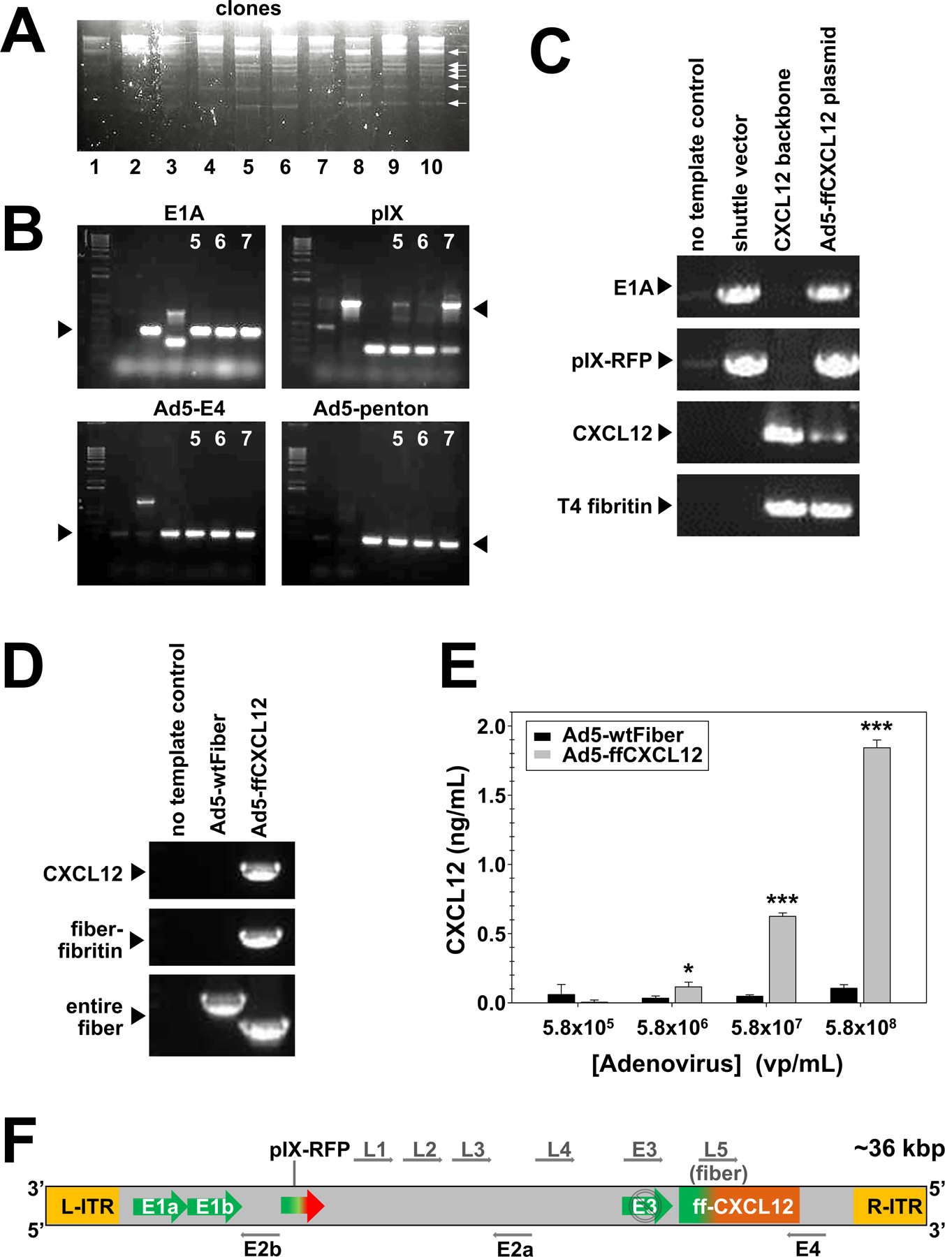
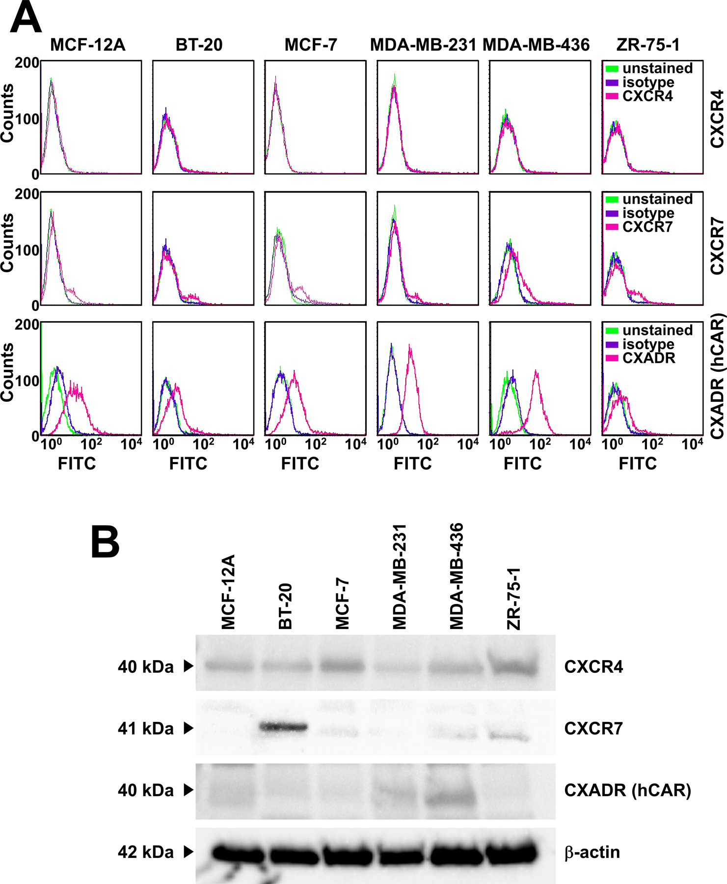
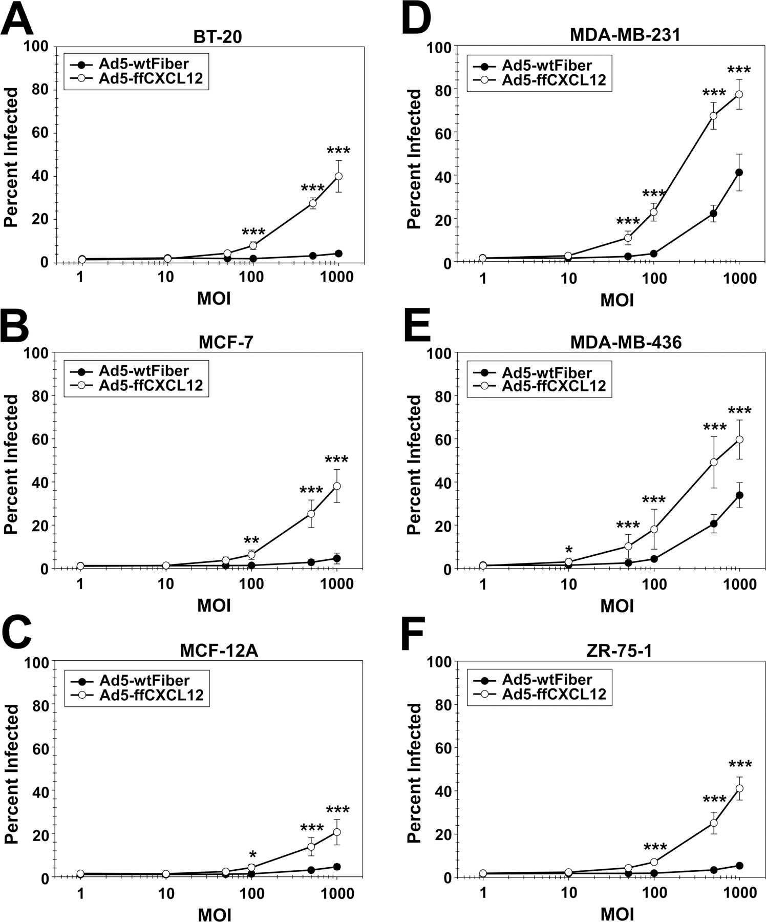
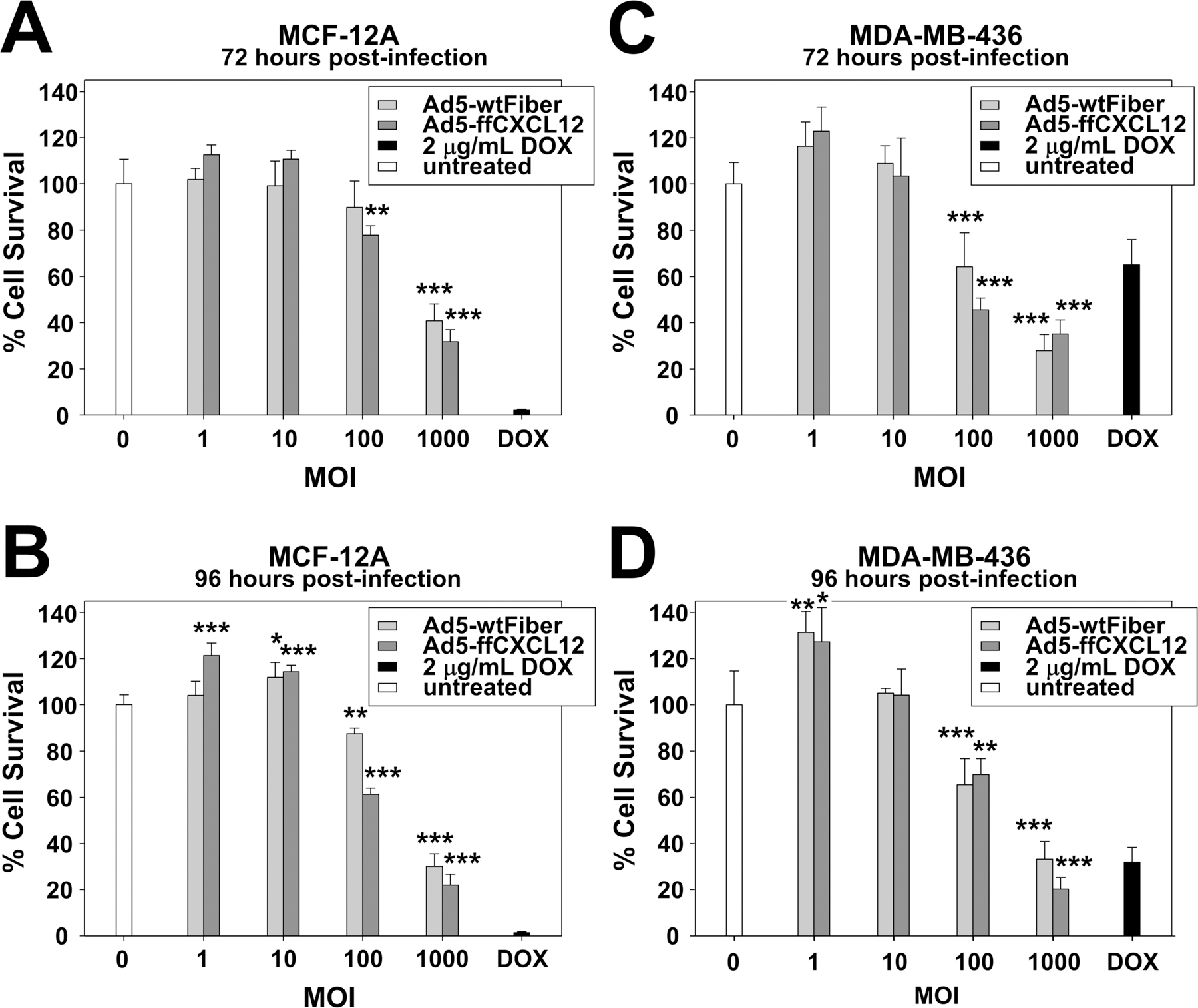
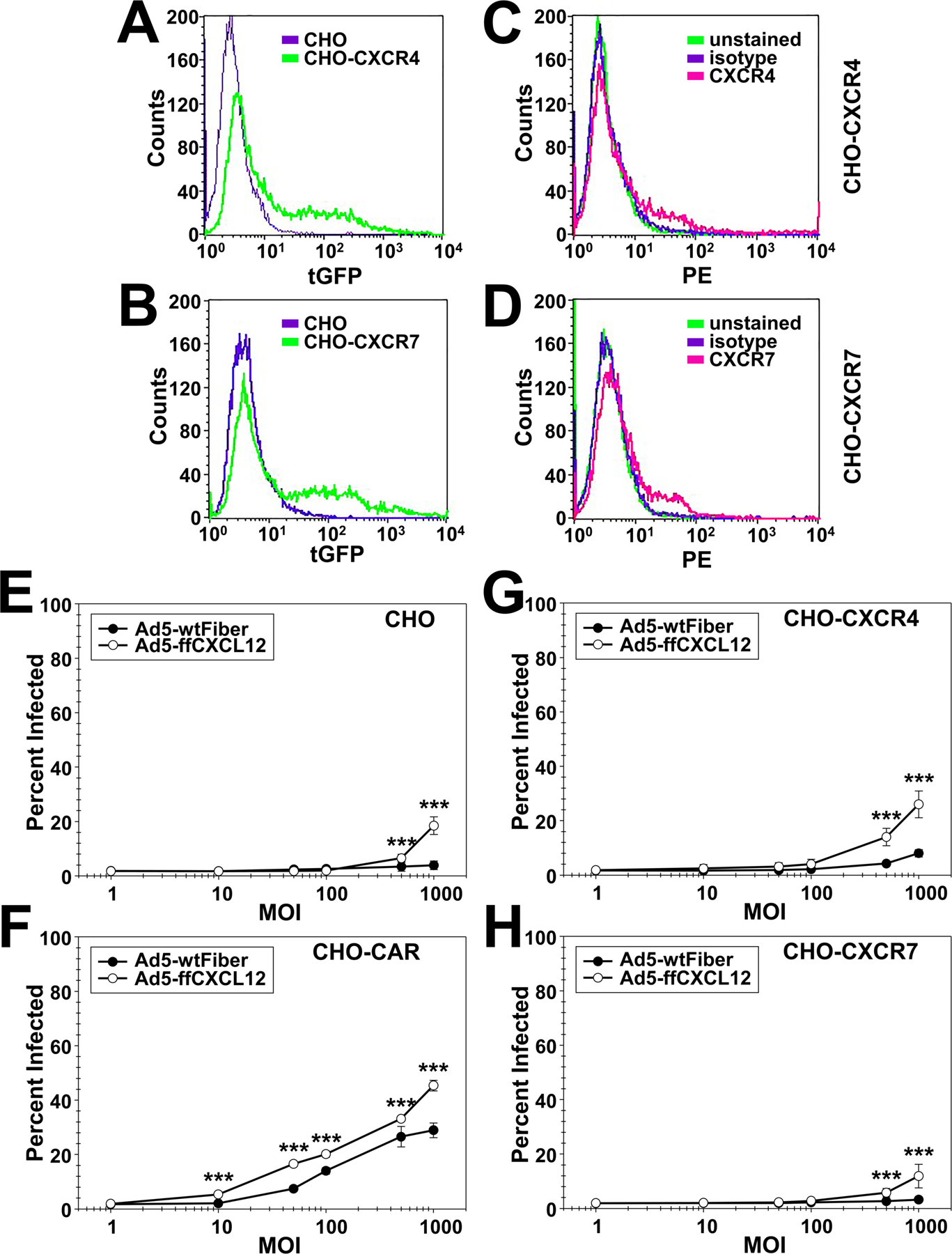
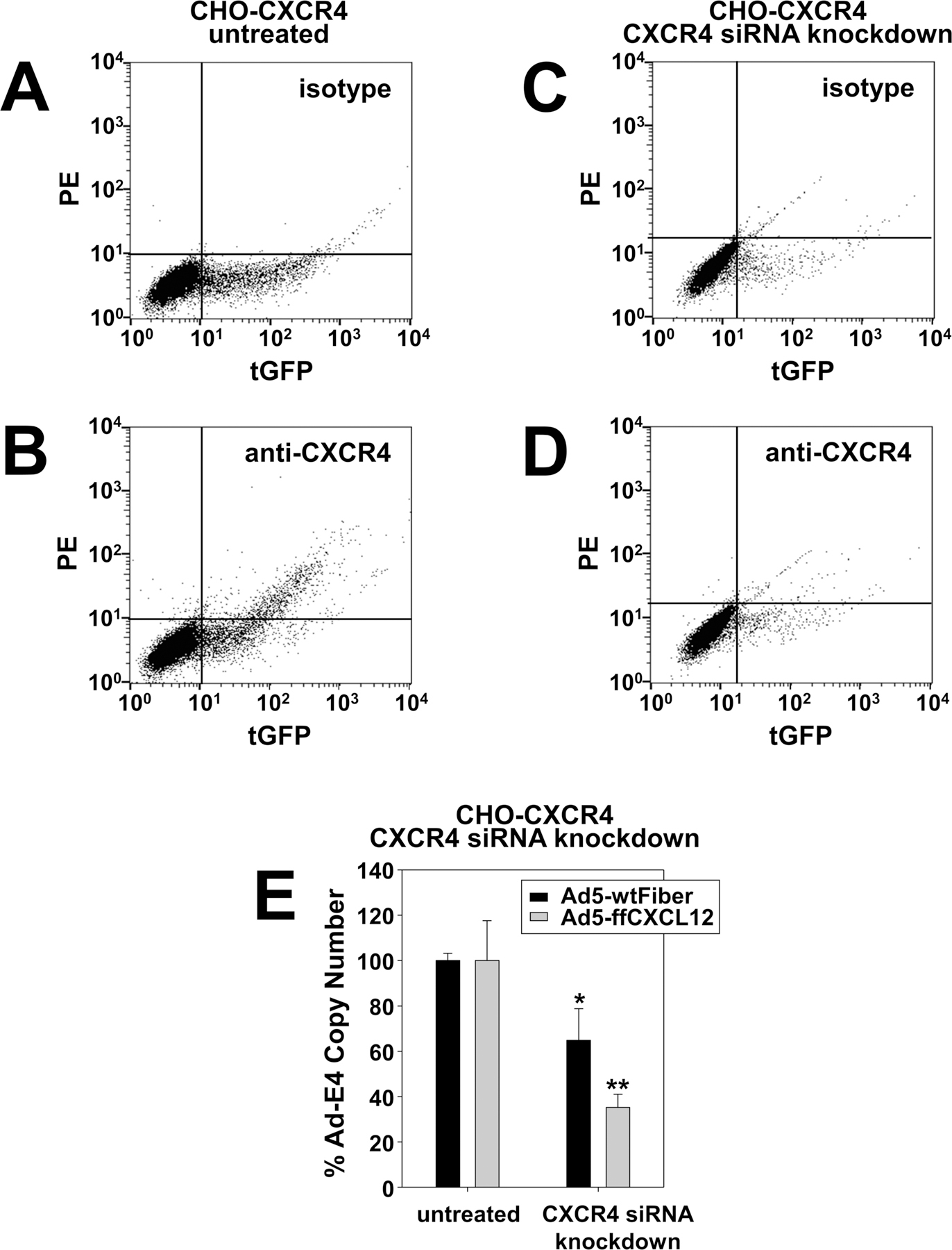
Breast cancer is the most frequently diagnosed cancer in women under 60, and the second most diagnosed cancer in women over 60. While significant progress has been made in developing targeted therapies for breast cancer, advanced breast cancer continues to have high mortality, with poor 5-year survival rates. Thus, current therapies are insufficient in treating advanced stages of breast cancer; new treatments are sorely needed to address the complexity of advanced-stage breast cancer. Oncolytic virotherapy has been explored as a therapeutic approach capable of systemic administration, targeting cancer cells, and sparing normal tissue. In particular, oncolytic adenoviruses have been exploited as viral vectors due to their ease of manipulation, production, and demonstrated clinical safety profile. In this study, we engineered an oncolytic adenovirus to target the chemokine receptors CXCR4 and CXCR7. The overexpression of CXCR4 and CXCR7 is implicated in the initiation, survival, progress, and metastasis of breast cancer. Both receptors bind to the ligand, CXCL12 (SDF-1), which has been identified to play a crucial role in the metastasis of breast cancer cells. This study incorporated a T4 fibritin protein fused to CXCL12 into the tail domain of an adenovirus fiber to retarget the vector to the CXCR4 and CXCR7 chemokine receptors. We showed that the modified virus targets and infects CXCR4- and CXCR7-overexpressing breast cancer cells more efficiently than a wild-type control vector. In addition, the substitution of the wild-type fiber and knob with the modified chimeric fiber did not interfere with oncolytic capability. Overall, the results of this study demonstrate the feasibility of retargeting adenovirus vectors to chemokine receptor-positive tumors.

Keywords: Adenovirus; Breast Cancer; CXCL12; CXCR4; CXCR7; Cancer; Chemokine; Oncolytic; Preclinical; Receptor; Virotherapy; Virus.


Conflict of interest statement

Conflicts of Interest The authors declare no conflict of interest. The funders had no role in the design of the study, in the collection, analyses, or interpretation of data, in the writing of the manuscript, or in the decision to publish the results.

Figures

Figure 1.
Figure 1.
Recombinant adenovirus clone confirmation. (A) DNA isolated from potential recombinant clones digested with Hind III. (B) Clones 5, 6, and 7 screened for E1A, pIX, Ad5-E4, and Ad5-penton by PCR. (C) PCR analysis of recombinant plasmid DNA from clone 7 by PCR. (D) PCR analysis of final virus stock after amplification in 293A-CXCR4 cell line and purification with CsCl gradient. (E) ELISA analysis of CXCL12 protein expression in purified viral stock. Bars are representative of mean values S.D. The differences between the two virus constructs at each adenovirus concentration were compared using Student’s t-test and were considered statistically significant if p < 0.05. *p < 0.05, ***p < 0.001. (F) Schematic of Ad5-ffCXCL12 genome. Abbreviations: VP, viral particles; PCR, polymerase chain reaction; CsCl, cesium chloride; S.D., standard deviation.
Figure 2.
Figure 2.
Expression of CXCR4 and CXCR7 in breast cancer cell lines. (A) Cell receptor expression was determined by flow cytometry of breast cancer cell lines immunostained with FITC-conjugated antibodies specific for CXCR4, CXCR7, and CXADR (a.k.a. CAR). Fluorescence detection of unstained cells (green peaks) was compared with cells immunostained with an isotype IgG staining (purple peaks) or a receptor-specific antibody (magenta peaks). In each experiment, 10,000 cells were analyzed for each sample. Shown are representative results of three independent experiments. (B) Western blot analysis of breast cancer cell lines. Aliquots of whole-cell lysates from each cell line were separated by SDS-PAGE, electroblotted onto nitrocellulose membranes, and probed with primary mouse anti-human antibodies specific for CXCR4 CXCR7, CXADR (a.k.a. CAR), or ?-actin. Membranes were subsequently incubated with a horseradish peroxidase-conjugated goat anti-mouse IgG antibody, developed using an ECL reagent, visualized by a Western blotting imaging system. Shown are representative blots after visualization. Abbreviations: ECL, enhanced chemiluminescent; FITC, fluorescein isothiocyanate; SDS-PAGE, sodium dodecylsulfate-polyacrylamide gel electrophoresis.
Figure 3.
Figure 3.
Detection of adenovirus infection. Percent of (A) BT-20, (B) MCF-7, (C) MCF-12A, (D) MDA-MB-231, (E) MDA-MB-436, and (F) ZR-75-1 cell lines infected with Ad5-wtFiber (●) or Ad5-ffCXCL12 (○) after treatment for 72 hours with increasing MOI was determined by flow cytometry analysis of pIX-RFP expression. All data is representative of three replicate experiments. Points indicate the mean S.D. of percent RFP positive cells. Two-way ANOVA followed by Bonferroni’s multiple comparisons test was performed to compare the percent infected cells at each MOI. The differences between the two virus treatments at each MOI were considered statistically significant if p < 0.05; *p < 0.05, **p < 0.01, ***p < 0.001. Abbreviations: MOI, multiplicity of infection; S.D., standard deviation.
Figure 4.
Figure 4.
Viability assays of MCF-12A and MDA-MB-436 after infection with increasing MOI of Ad5-wtFiber or Ad5-ffCXCL12. MCF-12A cells were infected with increasing MOI of Ad5-wtFiber or Ad5-ffCXCL12 for (A) 72 hours or (B) 96 hours before XTT assays. MDA-MB-436 cells were also infected with increasing MOI of Ad5-wtFiber or Ad5-ffCXCL12 for (C) 72 hours or (D) 96 hours before XTT assays. As a positive control, the cells were treated 2 ?g/mL DOX. All data are representative of five replicates normalized to untreated cells (100% cell viability). Bars indicate the mean S.D. Two-way ANOVA followed by Bonferroni’s multiple comparisons test was performed to compare the percent cell survival at each MOI to uninfected cells (0 MOI). The differences between the virus treatment at each MOI and uninfected cells were considered statistically significant if p < 0.05; *p < 0.05, **p < 0.01, ***p < 0.001. Abbreviations: DOX, doxorubicin; MOI, multiplicity of infection; S.D., standard deviation.
Figure 5.
Figure 5.
Expression of CXCR4 and CXCR7 in stably transfected CHO cells. GFP-tagged receptor expression in (A) CHO-CXCR4 and (B) CHO-CXCR7 cells was compared to parental CHO cells by flow cytometry after stable transfection with mammalian expression plasmids. CHO-CXCR4 and CHO-CXCR7 cells were also examined by immunostaining for cell surface expression of (C) CXCR4 or (D) CXCR7 using PE-conjugated monoclonal antibodies specific for the human receptors. The cells were incubated with PBS alone (unstained), an isotype control antibody, or the receptor-specific antibody. Following incubation, the cells were washed, resuspended in 0.4 mL PBS, and analyzed by flow cytometry. The percent of (E) CHO, (F) CHO-CAR, (G) CHO-CXCR4, and (H) CHO-CXCR7 cell lines infected with Ad5-wtFiber (●) or Ad5-ffCXCL12 (○) for 72 hours at increasing MOI was determined by flow cytometry analysis of pIX-RFP expression. All data is representative of three replicate experiments. Points indicate the mean S.D. of percent RFP positive cells. Two-way ANOVA followed by Bonferroni’s multiple comparisons test was performed to compare the percent infected cells at each MOI. The differences between the two virus treatments at each MOI were considered statistically significant if p < 0.05; ***p < 0.001. Abbreviation: MOI, multiplicity of infection; PE, phycoerythrin; RFP, red fluorescent protein; S.D., standard deviation; tGFP, turbo green fluorescent protein.
Figure 6.
Figure 6.
Effect of CXCR4 knockdown on adenovirus binding. Parental CHO-CXCR4 cells and CHO-CXCR4 cells transfected with CXCR4 siRNA were characterized by two parameter (dual-color fluorescence) flow cytometry. Shown are representative fluorescence plots of tGFP-tagged receptor expression (x axis) and immunostaining of CXCR4 expression (y axis) in untransfected cells using an isotype control (A) or a PE-conjugated monoclonal antibody specific for the human receptor (B). CXCR4 expression was also determined in cells transfected for 72 hours with an anti-CXCR4 siRNA. Shown are representative fluorescence plots of tGFP-tagged receptor expression (x axis) and immunostaining of CXCR4 expression (y axis) in transfected cells using an isotype control (C) or a PE-conjugated monoclonal antibody specific for the human receptor (D). Binding of Ad5-wtFiber (black bars) or Ad5-ffCXCL12 (grey bars) at 100 MOI was determined by qPCR of DNA isolated from untransfected CHO-CXCR4 cells or CHO-CXCR4 cells transfected for 72 hours with an anti-CXCR4 siRNA (E). All data are representative of five replicates normalized to untransfected cells (100% cell viability). Bars indicate the mean S.D. The differences between the untransfected and transfected groups were compared using Student’s t-test and were considered statistically significant if p < 0.05; *p < 0.05, **p < 0.01. Abbreviations: MOI, multiplicity of infection; PE, phycoerythrin; qPCR, quantitative polymerase chain reaction; S.D., standard deviation; siRNA, small interfering RNA; tGFP, turbo green fluorescent protein.

References

    1. Siegel RL, Miller KD and Jemal A (2019) Cancer Statistics, 2019. CA: A Cancer Journal for Clinicians, 69, 7–34. 10.3322/caac.21551 - DOI - PubMed
    1. O’Bryan SM and Mathis JM (2018) Oncolytic Virotherapy for Breast Cancer Treatment. Current Gene Therapy, 18, 192–205. 10.2174/1566523218666180910163805 - DOI - PMC - PubMed
    1. Aghi M and Martuza RL (2005) Oncolytic Viral Therapies—The Clinical Experience. Oncogene, 24, 7802–7816. 10.1038/sj.onc.1209037 - DOI - PubMed
    1. Alemany R, Balague C and Curiel DT (2000) Replicative Adenoviruses for Cancer Therapy. Nature Biotechnology, 18, 723–727. 10.1038/77283 - DOI - PubMed
    1. Alemany R (2014) Oncolytic Adenoviruses in Cancer Treatment. Biomedicines, 2, 36–49. 10.3390/biomedicines2010036 - DOI - PMC - PubMed

LinkOut - more resources