

Expression of autophagy and apoptosis-related factors in the periodontal tissue of experimental diabetic rats: a histomorphometric, microtomographic and immunohistochemical study
- PMID: 34178461
- PMCID: PMC8197035
- DOI: 10.7717/peerj.11577
Expression of autophagy and apoptosis-related factors in the periodontal tissue of experimental diabetic rats: a histomorphometric, microtomographic and immunohistochemical study
Abstract
Objective: This study aimed to investigate the expression of autophagy-related factors microtubule-associated protein l light chain 3 (LC3) and the apoptosis-related factors BCL2-associated X protein (Bax) and B cell lymphoma-2 (Bcl-2) in the periodontal tissue of experimental diabetic rats. These data were used to explore the potential mechanism in diabetes-induced periodontal tissue lesions.
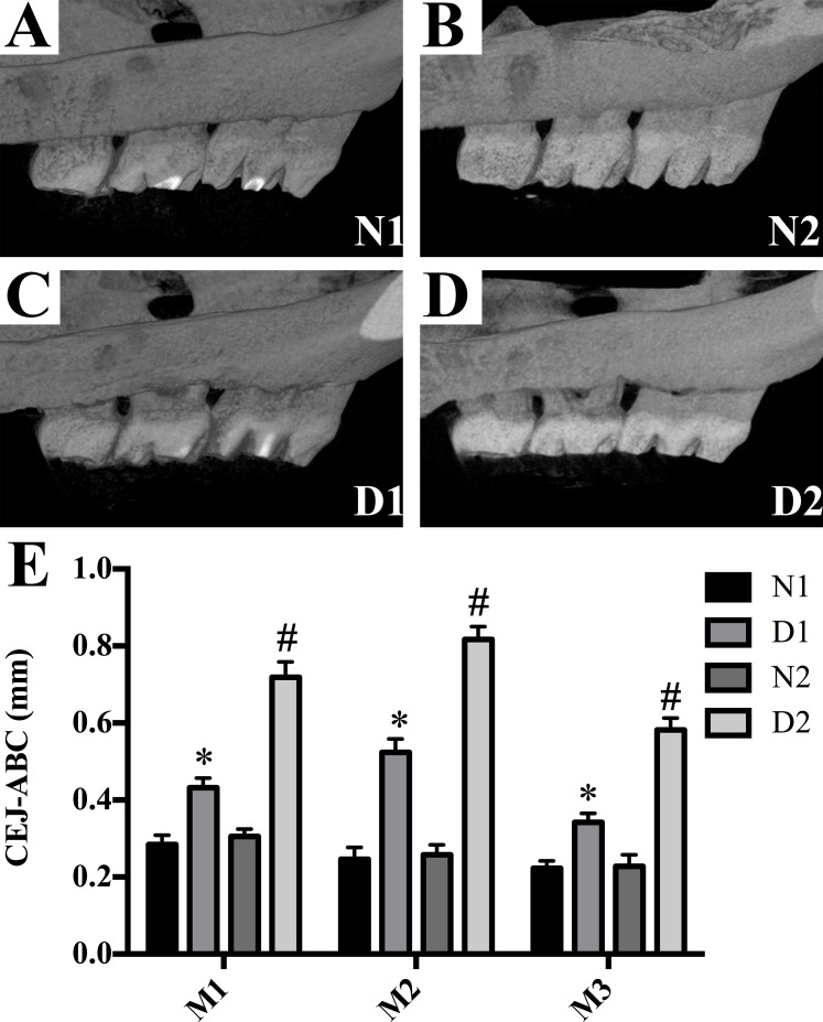
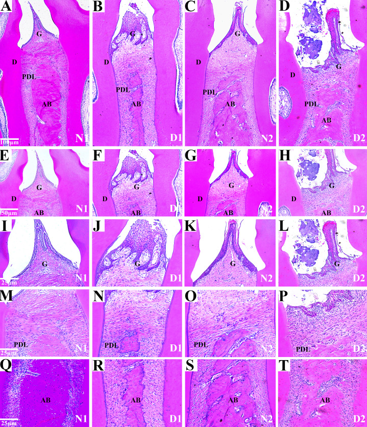
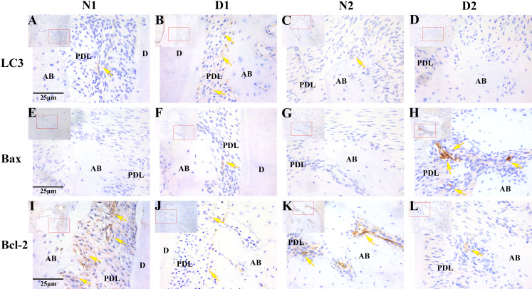
Methods: A total of 32 Sprague Dawley (SD) rats were randomly assigned into diabetes (group D, n = 16) and control groups (group N, n = 16). The diabetic group was induced by intraperitoneal injection of 1% streptozotocin (STZ, 60 mg/kg) and the control group was injected with citrate buffer (0.1mol/L). Rats were sacrificed after 4 and 8 weeks of feeding and collected as D1, N1 groups and D2, N2 groups, and the maxilla were retained for analysis. The changes in periodontal tissue structure were observed by hematoxylin-eosin (HE) staining. The expression and distribution of LC3, Bax and Bcl-2 in the periodontium of the rats was detected by immunohistochemical (SP) staining.
Results: Diabetic rats showed several changes compared to control animals including sparse alveolar bone trabecular structure, loss of the lamina dura and absorption of the local alveolar bone. The positive expression level of LC3 in the gingival epithelial, periodontal ligament and alveolar bone of group D1 was significantly higher than in the N1, N2 and D2 groups (P < 0.05). The level of Bax expression in the group D2 rats was significantly higher than those in the N1, N2 and D1 groups (P < 0.05), while the positive degree of Bcl-2 was significantly lower than those of other groups (P < 0.001). LC3 was negatively correlated with Bax and was irrelevant with Bcl-2; Bcl-2 was not correlated with Bax.
Conclusions: The expression of LC3, Bax and Bcl-2 changes in the periodontal tissue of diabetic rats may indicate that autophagy and apoptotic are involved in the process of periodontal tissue damage in diabetic rats. These changes may be one of the mechanisms of periodontal tissue lesions.
Keywords: Bax; Bcl-2; Diabetes; LC3; Periodontal tissue.
© 2021 Mei et al.
Conflict of interest statement
The authors declare that they have no competing interests.
Figures
References
-
- Alikhani Z, Alikhani M, Boyd CM, Nagao K, Trackman PC, Graves DT. Advanced glycation end products enhance expression of pro-apoptotic genes and stimulate fibroblast apoptosis through cytoplasmic and mitochondrial pathways. Journal of Biological Chemistry. 2005;280(13):12087–12095. doi: 10.1074/jbc.M406313200. - DOI - PubMed
-
- Balci Yuce H, Karatas O, Aydemir Turkal H, Pirim Gorgun E, Ocakli S, Benli I, Cayli S. The effect of melatonin on bone loss, diabetic control, and apoptosis in rats with diabetes with ligature-induced periodontitis. Journal of Periodontology. 2016;87(4):e35–e43. doi: 10.1902/jop.2015.150315. - DOI - PubMed
-
- Bascones-Martinez A, Gonzalez-Febles J, Sanz-Esporrin J. Diabetes and periodontal disease. Review of the literature. American Journal of Dentistry. 2014;27(2):63–67. - PubMed
-
- Bullon P, Cordero MD, Quiles JL, del Carmen Ramirez-Tortosa M, Gonzalez-Alonso A, Alfonsi S, García-Marín R, De Miguel M, Battino M. Autophagy in periodontitis patients and gingival fibroblasts: unraveling the link between chronic diseases and inflammation. BMC Medicine. 2012;10(1):122. doi: 10.1186/1741-7015-10-122. - DOI - PMC - PubMed
LinkOut - more resources
Full Text Sources
Research Materials
