

Non-Compressive, Disabling, Cervical Radiculopathy and Neck Pain: Cave Osteoid Osteoma
- PMID: 34178528
- PMCID: PMC8221648
- DOI: 10.7759/cureus.15209
Non-Compressive, Disabling, Cervical Radiculopathy and Neck Pain: Cave Osteoid Osteoma
Abstract
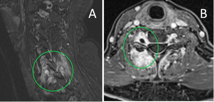
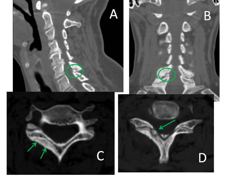
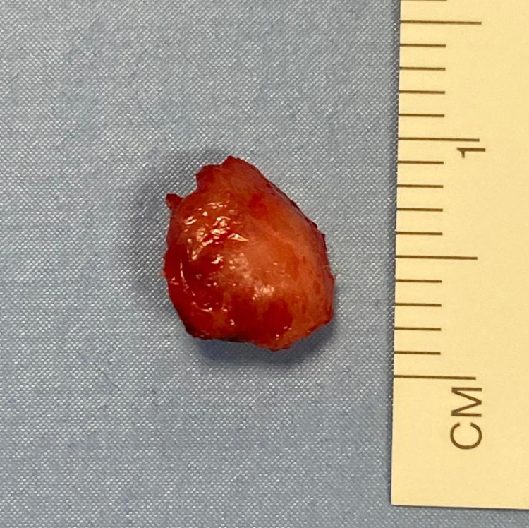
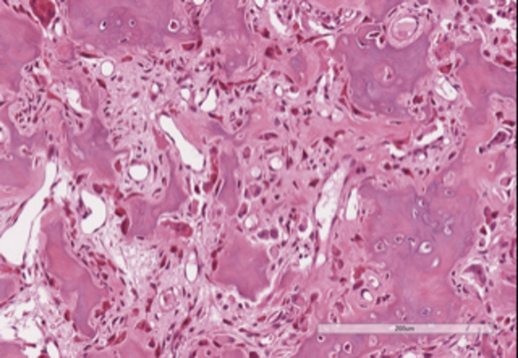
Cervical radiculopathy is a common clinical condition with an annual incidence of 85/10,000. Refractory cases with positive disco-vertebral imaging findings are routinely referred to the Neurosurgeon for evaluation and treatment. In the absence of a clearcut compressive etiology, other rarer but surgically curable causes must be considered before recommending conservative management. We discuss the case of an otherwise active, healthy patient with an invalidating, refractory, relapsing nuchal pain and cervical radiculopathy. Only careful and state-of-the-art neuroimaging led to the correct diagnosis: an osteoid osteoma of the right C6 lamina was diagnosed and microsurgically resected allowing complete recovery and cure. The clinical features of these rare tumors in this unusual location are reviewed. The case is relevant for multifold reasons: it draws attention to rare conditions which can mimic radicular compression; emphasizes the need for a careful evaluation and appreciation of specific clinical symptoms and signs associated with non-compressive radiculopathies; prompts planning of a state of the art imaging workup, in order to rule out such an elusive tumor. All these measures minimize the risk of overlooking the present and other rare pathologies, sparing patients a long path of time-consuming, frustrating and cost-ineffective studies and treatment modalities.
Keywords: bone tumors; general neurosurgery; neuro mri; ortho surgery; primary spine tumour; spinal pain.
Copyright © 2021, Brunori et al.
Conflict of interest statement
The authors have declared that no competing interests exist.
Figures
References
-
- Cervical radiculopathy: pathophysiology, presentation, and clinical evaluation. Abbed KM, Coumans JV. Neurosurgery. 2007;60:0–34. - PubMed
-
- Incidence and epidemiology of cervical radiculopathy in the United States military: 2000 to 2009. Schoenfeld AJ, George AA, Bader JO, Caram PM Jr. http://10.1097/BSD.0b013e31820d77ea. J Spinal Disord Tech. 2012;25:17–22. - PubMed
-
- Prevalence of cervical spondylotic radiculopathy: a door-to-door survey in a Sicilian municipality. Salemi G, Savettieri G, Meneghini F, et al. Acta Neurol Scand. 1996;93:184–188. - PubMed
-
- Cervical radiculopathy: epidemiology, etiology, diagnosis, and treatment. Woods BI, Hilibrand AS. J Spinal Disord Tech. 2015;28:0–9. - PubMed
Publication types
LinkOut - more resources
Full Text Sources