Review
. 2021;136(6):675.
doi: 10.1140/epjp/s13360-021-01657-9. Epub 2021 Jun 20.

Role of plasmonics in detection of deadliest viruses: a review

Affiliations
Review

Role of plasmonics in detection of deadliest viruses: a review

Foozieh Sohrabi et al. Eur Phys J Plus. 2021.

Abstract

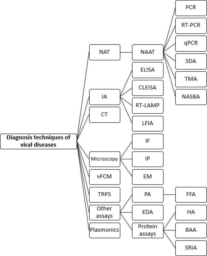
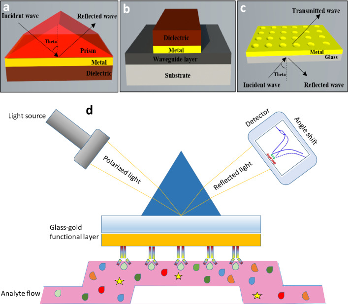
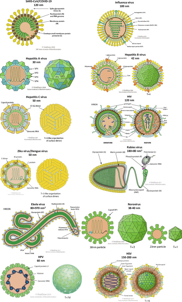
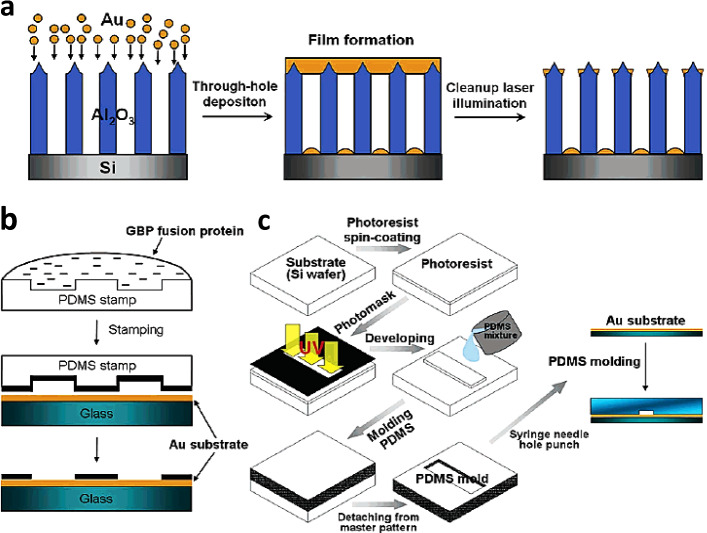
Viruses have threatened animal and human lives since a long time ago all over the world. Some of these tiny particles have caused disastrous pandemics that killed a large number of people with subsequent economic downturns. In addition, the quarantine situation itself encounters the challenges like the deficiency in the online educational system, psychiatric problems and poor international relations. Although viruses have a rather simple protein structure, they have structural heterogeneity with a high tendency to mutation that impedes their study. On top of the breadth of such worldwide worrying issues, there are profound scientific gaps, and several unanswered questions, like lack of vaccines or antivirals to combat these pathogens. Various detection techniques like the nucleic acid test, immunoassay, and microscopy have been developed; however, there is a tradeoff between their advantages and disadvantages like safety in sample collecting, invasiveness, sensitivity, response time, etc. One of the highly resolved techniques that can provide early-stage detection with fast experiment duration is plasmonics. This optical technique has the capability to detect viral proteins and genomes at the early stage via highly sensitive interaction between the biological target and the plasmonic chip. The efficiency of this technique could be proved using commercialized techniques like reverse transcription polymerase chain reaction (RT-PCR) and enzyme-linked immunosorbent assay (ELISA) techniques. In this study, we aim to review the role of plasmonic technique in the detection of 11 deadliest viruses besides 2 common genital viruses for the human being. This is a rapidly moving topic of research, and a review article that encompasses the current findings may be useful for guiding strategies to deal with the pandemics. By investigating the potential aspects of this technique, we hope that this study could open new avenues toward the application of point-of-care techniques for virus detection at early stage that may inhibit the progressively hygienic threats.


Figures

Fig. 1
Fig. 1
Detection techniques for viral diseases. NAT Nucleic Acid Test, IA Immunoassay, CT Computed tomography, vFCM viral Flow Cytometry, TRPS Tunable resistive pulse sensing, NAAT Nucleic Acid Amplification Test, ELISA enzyme-linked immunosorbent assay, CLEIA Chemiluminescence enzyme immunoassay, RT-LAMP reverse transcription loop-mediated isothermal amplification assay, LFIA lateral flow immunochromatographic assays, IF Immunofluorescence, IP Immunoperoxidase, EM Electron Microscopy, PA Plaque assay, EDA Endpoint Dilution Assay, PCR Polymerase Chain Reaction, RT-PCR Reverse Transcription-PCR, qPCR quantitative PCR, SDA strand displacement assay, TMA Transcription-Mediated Assay, NASBA Nucleic acid sequence-based amplification (NASBA), FFA Focus forming assay (FFA), HA Hemagglutination Assay, BAA Bicinchoninic Acid Assay, SRIA Single Radial Immunodiffusion Assay
Fig. 2
Fig. 2
Coupling methods of incident light to SPs a Kretschmann−Raether arrangement (prism coupling) b waveguide coupling c plasmonic crystal (grating coupling) [18]. d Schematic of the conventional plasmonic biosensor with immobilized antibodies on the gold surface. Binding of the antigens causes a difference in SPR signal [35]
Fig. 3
Fig. 3
The schematics of the viruses, their size, and structure that are studied in the current review. The virus schematics are extracted from the website of Swiss Institute of Bioinformatics [42]. These viruses are SARS-CoV/COVID-19, Influenza, Hepatitis A, Hepatitis B, Hepatitis C, HIV, Zika, Dengue, Rabies, Ebola, Norovirus, HPV and HSV with the diameters of 120 nm, 100 nm, 30 nm, 42 nm, 50 nm, 120 nm, 50 nm, 50 nm, 180 × 80 nm2, 80 × 970 nm2, 38–40 nm, 60 nm and 150–200 nm
Fig. 4
Fig. 4
a Laser-ablated deposition method used by Park et al. [54] for the generation of gold nanopatterns b microcontact printing procedure using the PDMS stamp, and c the fabrication process of the PDMS microfluidic device by photolithography. This chip was used as a sensing platform for SARS CoV
Fig. 5
Fig. 5
Influenza A and B are most common influenza types for human. Influenza A is divided into two main subgroups of A (H1N1) and A (H3N2) and influenza B has two lineages of B (Victoria) and B (Yamagata) [146]
Fig. 6
Fig. 6
a Schematic of the plasmonic chip used for the detection of H7N9 virus. The functionalized chip surface provided the selective binding for H7N9 by Chang et al. [77]. b The selection and application of a cognate pair of aptamers for H5N2 avian influenza. Kim et al. [78] have applied graphene-oxide-based systemic evolution of ligands by exponential enrichment (GO-SELEX). They have found that the aptamers of J3APT and JH4APT played the role of a cognate pair which bound to the virus at different locations simultaneously [78]. c The SPR optical fiber sensor surface was modified with the carboxyl group SAM layer and then activated by EDC/NHS. Zhao et al. [80] have proposed this optical fiber sensor for the detection of influenza H6 virus. d The aggregation of the glyconanoparticles in the presence of the influenza virus. At the presence of the influenza virus, the solution of the glyconanoparticles aggregated that caused a color change within 30 min [82]. e The gold binding polypeptide (GBP)-fusion protein was immobilized on the gold surface that could provide interaction with AIa and its anti-AI antibody [83]
Fig. 7
Fig. 7
A plasmon-assisted fluoro-immunoassay (PAFI) for detection of influenza virus using gold NPs decorated with carbon nanotubes (AuCNTs) a Preparation of AuNP-decorated CNT nanostructures (AuCNTs) b the process of influenza virus detection by using PAFI, non-scalable [86]
Fig. 8
Fig. 8
a Preparation Steps and SEF/Protease-Based Sandwich-Type Immunoassay for Hepatitis B Detection. Ghafary et al. [87] have used these NPs as a fluorescence label where the plasmonic phenomenon amplified the fluorescence intensity. b The sandwich structure of Ag nanorice@Raman label@SiO2 and the operating principle of the SERS sensor for HBV DNA detection. Li et al. [90] have introduced plasmonic nanorice antenna on triangle nanoarray for surface-enhanced Raman scattering detection of hepatitis B virus DNA. c A biosensor for hepatitis B virus detection using GBP fusion proteins proposed by Zheng et al. [91]
Fig. 9
Fig. 9
a Synthesis and chemical structure of the poly(HPMA-co-CBMAA) polymer brush (left) and schematics of the gold surface with the tethered brush that is post-modified with protein ligand (right). b Riedel et al. [92] have proposed the SPR setup and sensor chip with poly(HPMA-co-CBMAA) brush functioning as a binding matrix for direct detection of the anti-HBs target analyte
Fig. 10
Fig. 10
Plasmon-enhanced fluorescence spectroscopy biosensor with detail of sensor chip with poly(HPMA-co-CBMAA) brush functioning as a binding matrix [93]
Fig. 11
Fig. 11
Schematic of HIV genome structure [186]
Fig. 12
Fig. 12
Structure of an HIV virion particle on left image a and, electron microscope (issued by CDC Public Health Image Library) on right image b. Immunoblot bands on the left side of image b are showing Gp Glycoprotein, p protein, SU surface protein, TM transmembrane protein, gp120 (precursor of SU and TM), RT reverse transcriptase, IN integrase, CA capsid protein, MA matrix protein, PR protease, NC nucleic acid binding protein, LI link protein. MHCs (major histocompatibility complexes) are HLA antigens. Table 1 explicitly discusses the function of the different proteins [186]. (Graphic Hans Gelderblom, Robert Koch Institute, Berlin)
Fig. 13
Fig. 13
Cyclic conversion of immature HIV to mature HIV
Fig. 14
Fig. 14
Schematic of stepping bio-functioning process of the sandwich assay. a Top schematic, the functionalized top surface of cantilever treated by captured antibodies used against HIV-1 p24 antigen. Middle schematic, immersing the cantilever in the human serum sample provides the condition of binding to p24 to the cantilever surface. Bottom schematic, trapping 100-nm-dimeter gold NPs by the p24 antigen. b Scanning electron microscopy (SEM) image of the silicon micro-cantilever arrays c. 96-well microtiter plate schematic used for immunoassays experiment [94]
Fig. 15
Fig. 15
Immunoreaction response to nanomechanical and optoplasmonic transduction mechanisms. a Schematic to the demonstration of simultaneous optical beam deflection and microcantilever vibration. A laser beam is focused onto the cantilever’s reed, and at the same time, the deflection of the reflected beam during vibration by a piezoelectric actuator beneath the micro-cantilever array chip measured using a sensitive photodetector positioned on the top surface of micro-cantilever. b Resonance frequency peaks (bottom graphs) of the first three vibration modes (top panels) of a silicon cantilever before (dotted lines) and after (solid lines) the immunoreaction assay for a blank human serum sample (control) and 5 × 10 − 4 pg/mL of p24 in human serum. The relative variations of the resonance frequencies are shown beside the resonance frequency peaks. c Schematics of the multiple pathways for light scattering by the gold NPs bound to the micro-cantilever. d Darkfield optical images of the microcantilevers analyzed in b after the immunoassays. The images show the microcantilever and chip preclamping regions. At the right, zoomed images of regions at the preclamping and the microcantilever are also shown [94]
Fig. 16
Fig. 16
a Overall description of BPD biosensor. a and b The process for elicitation of immune response by functionalization of AuNRs with ZIKV-NS1 protein [97]. b Consequences of assemblage of ox-GCE-[AuNPs-SiPy]/ZIKV1 biosensor and DNA hybridization for ZIKV detection [99]. c Schematic representation of the stages of biosensor construction and drug delivery [101]
Fig. 17
Fig. 17
A Representation of overall mechanism for RVG-PEG-AuNRs@SiO2 delivery through neural passways and photothermal therapy. B Scheme steps for synthesis of Rabies virus-like silica-coated gold nanorods (RVG-PEG-AuNRs@SiO2): (a) Au seed, (b) longitudinal growth of AuNRs, (c) AuNRs after both of longitudinal and transverse growth, (d) silica-coated AuNRs and (e) RVG29 peptide-PEG5k-conjugated AuNRs@SiO2 (The total dimensions of the structure is identical to the Rabies virus (length/width ≅ 2.4)). C Morphological TEM images of steps (a-c and e) and gold NPs (RVG-PEG-AuNPs@SiO2; ≈68 000 nm3; f) [102]
Fig. 18
Fig. 18
a Stages for construction of NOV detection biosensor by use of hybrid nanocomposite [107]. b Schematic representing the aptamer-aptamer sandwich assay for trapping NOV capsid protein [109]
Fig. 19
Fig. 19
a (Left) Generally fabricated homogeneous and a heterogeneous assay. (Right) immobilization of AuNPs and UNCPs on the NAAO membrane [114]. b Schematic diagram of the process for multiple DNA detection. c First row: Represents SEM images of the conjunction of PNCs and magnetic beads in the presence of EBOV, VV, and BA, respectively. Second row: the magnified images of the first row[116]
Fig. 20
Fig. 20
a, b schematic showing three-dimensional of optofluidic nanoplasmonic biosensors at the absence and presence of captured virus at the surface of biosensor. c Detection of PT- EBOVs according to light transmission at concentration of 108 PFU/mL. d Repeatability of the spectral measurements with various biosensors [112]
Fig. 21
Fig. 21
a Optical micrographs (20x) taken from the DENV sample and Dengue particles plus AH peptide. The prior visualize light scattering from each DENV particles in the sample and the latter AH peptide rupture Dengue particles from the specimen. b The capture of virus particles and lipid vesicles by plasmonic nanohole array [118]. c The needful steps for the manufacture of the AuNP-E vaccine and its mechanism for serotype-specific neutralization of the DENV [120]
Fig. 22
Fig. 22
Human papillomavirus (HPV) infection and replication in cervical epithelial cells. a The normal cervix has a (narrow) transformation zone in which there is an abrupt transition from a columnar epithelium (sometimes via a metaplastic epithelium) to a squamous epithelium; HPVs are probably most infectious to cells that are close to this junction. b HPV viruses gain access to the basal epithelial cells of the cervix via the vagina (for example, during sexual intercourse), where they replicate episomally (outside the host chromosome in the cytoplasm) and express the (early) viral genes E1, E2, E4, E5, E6 and E7. c The infected basal cells, which show signs of cell disruption as a result of the viral infection, continue their differentiation and migration to the epithelial surface, where d the (now) squamous cells start to express the late HPV genes LI and L2. Infectious virus particles are formed and shed into the lumen of the vagina. (e) HPV infection (particularly with the high-risk types) can progress to: (1) HPV-induced mild dysplasia, (2) the final stages of cervical intraepithelial neoplasia (CIN3) and, eventually, (3) invasive cervical cancer (CaCx), when the basement membrane is breached by the cells, allowing local spread and also distant metastasis. (f) In transformed epithelial cells, HPV genes are integrated into the host chromosomes, with an expression of (the oncogenic) E6 and E7 proteins, which bind to the tumor-suppressor proteins p53 and Rb [14]
Fig. 23
Fig. 23
Schematic representation of Plasmonic quantum probe (PQPs) synthesis by forming an ion zone and HRTEM image of gold probes on silicon carrier [123]
Fig. 24
Fig. 24
a The Bio-TEM image showing the presence of PQPs inside the cancerous cells (HeLa), b Bio TEM images show the interaction between plasmonic probes and nucleus components. c Plasmonic Raman signals response for cervical cancer (HeLa) after 6 and 18 h of cell seeding to monitor the growth of cells by studying the G0 G1 and G3 phase of the cell cycle [123]
Fig. 25
Fig. 25
a Crystal structure of the HSV-1 gD protein in a free and synthetic body. (b) Overall monitoring of inhibitors that interfere with interactions between HSV‐1 gD and human receptors (HVEM or Nectin1) by SPR analysis [127]. c Schematic representation of selected NPAuG1-S2, NPAuG2-S4, and NPAuG3-S8 with various structures. d NPAuG3-S8 accumulation in cerebral tissue and time-dependence of crossing NPAuG3-S8 from BBB [129]

Similar articles

Cited by

References

    1. Organization, W.H.; Available from: https://www.who.int/emergencies/diseases/novel-coronavirus-2019
    1. Worldometer. Available from: https://www.worldometers.info/coronavirus/
    1. Rusnati M, et al. Bridging the past and the future of virology: surface plasmon resonance as a powerful tool to investigate virus/host interactions. Crit. Rev. Microbiol. 2015;41(2):238–260. doi: 10.3109/1040841X.2013.826177. - DOI - PubMed
    1. B. Marintcheva, Chapter 1 - Introduction to Viral Structure, Diversity and Biology∗∗Parts of this chapter were originally published in Marintcheva B. A box of paradoxes: the fascinating world of viruses. Bridgew Rev 2013;32(2):25–8. http://vc.bridgew.edu/br_rev/vol32/iss2/8 and are reproduced here with the permission of the editor, in Harnessing the Power of Viruses, B. Marintcheva, Editor. 2018, Academic Press. p. 1–26
    1. Huang JC, et al. Detection of severe acute respiratory syndrome (SARS) coronavirus nucleocapsid protein in human serum using a localized surface plasmon coupled fluorescence fiber-optic biosensor. Biosens. Bioelectron. 2009;25(2):320–325. doi: 10.1016/j.bios.2009.07.012. - DOI - PMC - PubMed

LinkOut - more resources