. 2021;9(5):539-545.
doi: 10.1007/s40336-021-00441-0. Epub 2021 Jun 18.

Ultrasound and shear-wave elastography patterns of COVID-19 mRNA vaccine-related axillary, supra and subclavicular lymphadenopathy

Affiliations

Ultrasound and shear-wave elastography patterns of COVID-19 mRNA vaccine-related axillary, supra and subclavicular lymphadenopathy

Divina D'Auria et al. Clin Transl Imaging. 2021.

Abstract

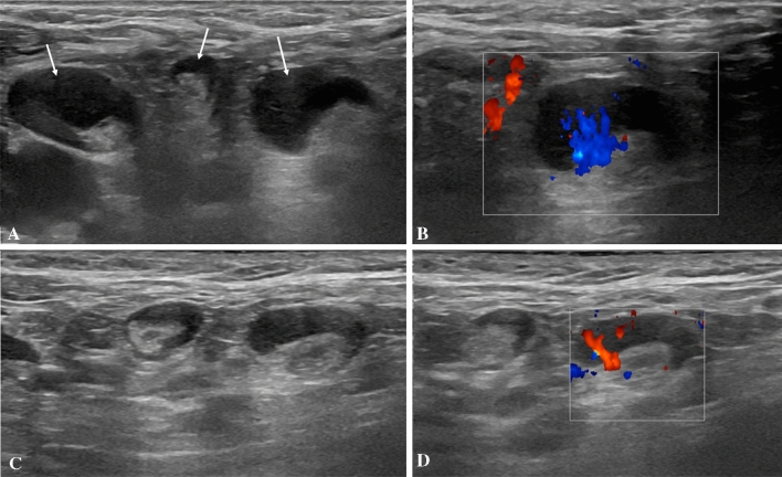
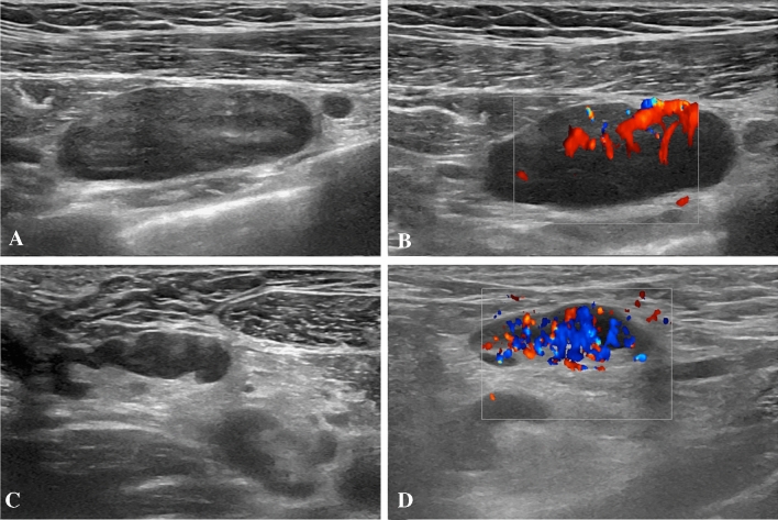
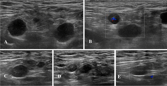
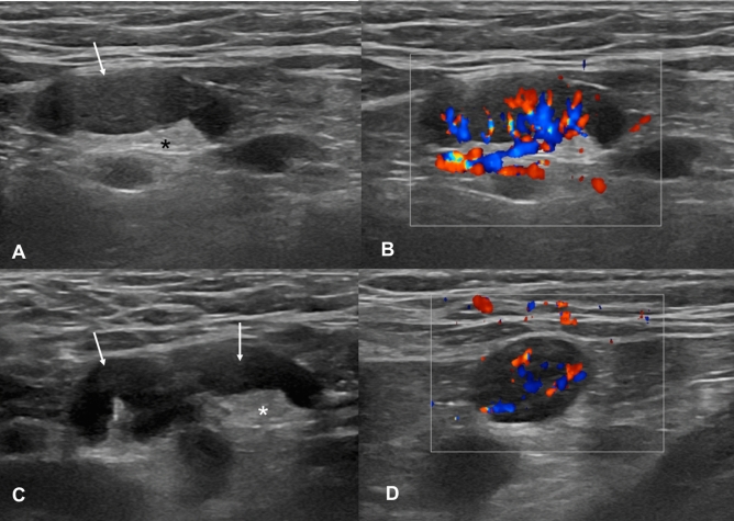
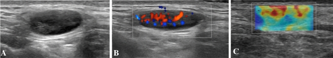
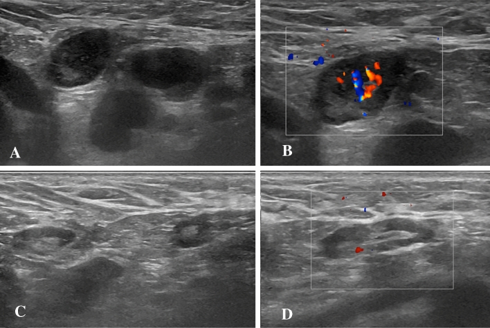
In this pictorial essay, we illustrate the ultrasound appearance of COVID-19 Pfizer-BioNTech vaccine-related lymph node abnormalities, which can occur at different stations ipsilateral to the site of vaccination, after either first or second vaccine dose and can represent a diagnostic dilemma when encountered in patients with underlying conditions. Typically, they appear as enlarged hypoechoic nodes with loss of fat hilum, increased hilar and cortical vascularization at color-Doppler, but low to intermediate cortical consistence at shear-wave elastography. Asymmetric or diffuse cortical thickening is also frequently encountered. They can be observed in patients without and with clinical symptoms, such as armpit pain, fever and fatigue.

Keywords: Covid-19; Lymphadenopathy; Shear-wave elastography; Ultrasound; Vaccine; mRNA vaccine.


Conflict of interest statement

Conflict of interestAll the authors declare that they have no conflict of interest.

Figures

Fig. 1
Fig. 1
B-Mode (A) and color-Doppler (B) US examination performed in a 28-year-old woman 2 days after receiving the second dose of vaccine: axillary lymph nodes (maximum diameter 14 × 9 mm) with fatty hilum but diffuse cortical thickening (maximum 10 mm, arrows in A) and vivid hilar and cortical vascularization (B) were detected. B-Mode (C) and color-Doppler (D) US examination performed after 4 weeks: reduction of lymph nodes size (7 × 6 mm), cortical thickness (2.9 mm) and vascularization was observed
Fig. 2
Fig. 2
B-Mode (A, B) and color-Doppler (B, D) US examination performed in a 28-year-old woman 2 days after receiving the second dose of vaccine. Enlarged (33 × 11 mm) subclavicular lymph node with no detectable fatty hilum (A) and vivid hilar and cortical vascularization (B) was detected. Enlarged axillary lymph node (26 × 7 mm) with cortical thickening (5 mm) (C) and increased vascularization (D) were also found
Fig. 3
Fig. 3
B-Mode (A) and color-Doppler (B) US examination performed 3 days after receiving the second vaccine dose. Multiple round (maximum diameter 11 × 10 mm) axillary lymph nodes with no fatty hilum and mild hilar vascularization were detected. B-Mode (C, D) and color-Doppler (E) US examination performed after 5 weeks. Only a reduction of lymph node size (maximum diameter 9 × 6 mm) was detectable.
Fig. 4
Fig. 4
B-Mode (A, C) and color-Doppler (B, D) US examination performed 3 days after receiving the second dose of vaccine. Enlarged (maximum diameter 21 × 7 mm) axillary lymph node with preserved fatty hilum (* in A and C), diffuse cortical thickening (6 mm, arrows in A and C) and vivid hilar and cortical vascularization (B, D) were found
Fig. 5
Fig. 5
B-Mode (A), color-Doppler (B) and shear wave (C) US examination performed 8 days after receiving the first dose of vaccine. Enlarged (14 × 6 mm) supraclavicular lymph node with no fatty hilum (A), increased hilar and cortical vascularization (B) and soft-intermediate consistence at shear-wave elastography (C) is shown
Fig. 6
Fig. 6
B-Mode (A) and color-Doppler (B) US examination performed 2 days after receiving the first dose of vaccine: multiple axillary lymph nodes (maximum diameter 15 × 10 mm) with poorly represented fatty hilum (A) and moderate vascularization (B) were found. B-Mode (C) and color-Doppler (D) US examination performed after 4 weeks. Although lymph node size remained stable, fatty hilum was restored (C) and vascularization reduced at color-Doppler (D)

References

    1. Dagan N, Barda N, Kepten E, Miron O, Perchik S, Katz MA, Hernán MA, Lipsitch M, Reis B, Balicer RD. BNT162b2 mRNA Covid-19 vaccine in a nationwide mass vaccination setting. N Engl J Med. 2021 doi: 10.1056/NEJMoa2101765. - DOI - PMC - PubMed
    1. Polack FP, Thomas SJ, Kitchin N, Absalon J, Gurtman A, Lockhart S, Perez JL, Pérez Marc G, Moreira ED, Zerbini C, Bailey R, Swanson KA, Roychoudhury S, Koury K, Li P, Kalina WV, Cooper D, Frenck RW, Jr, Hammitt LL, Türeci Ö, Nell H, Schaefer A, Ünal S, Tresnan DB, Mather S, Dormitzer PR, Şahin U, Jansen KU, Gruber WC, C4591001 Clinical Trial Group Safety and efficacy of the BNT162b2 mRNA Covid-19 vaccine. N Engl J Med. 2020;383(27):2603–2615. doi: 10.1056/NEJMoa2034577. - DOI - PMC - PubMed
    1. Becker AS, Perez-Johnston R, Chikarmane SA, Chen MM, El Homsi M, Feigin KN, Gallagher KM, Hanna EY, Hicks M, Ilica AT, Mayer EL, Shinagare AB, Yeh R, Mayerhoefer ME, Hricak H, Vargas HA. Multidisciplinary recommendations regarding post-vaccine adenopathy and radiologic imaging: radiology scientific expert panel. Radiology. 2021;24:210436. doi: 10.1148/radiol.2021210436. - DOI - PMC - PubMed
    1. Özütemiz C, Krystosek LA, Church AL, Chauhan A, Ellermann JM, Domingo-Musibay E, Steinberger D. Lymphadenopathy in COVID-19 vaccine recipients: diagnostic dilemma in oncology patients. Radiology. 2021;24:210275. doi: 10.1148/radiol.2021210275. - DOI - PMC - PubMed
    1. Ahn RW, Mootz AR, Brewington CC, Abbara S. Axillary lymphadenopathy after mRNA COVID-19 vaccination. Radiol Cardiothorac Imaging. 2021;3(1):e210008. doi: 10.1148/ryct.2021210008. - DOI - PMC - PubMed

LinkOut - more resources