

Case Report: Report of 2 Different Cases of Ovarian Teratoma Evaluated by Dynamic Contrast-Enhanced Ultrasound
- PMID: 34178898
- PMCID: PMC8226026
- DOI: 10.3389/fped.2021.681404
Case Report: Report of 2 Different Cases of Ovarian Teratoma Evaluated by Dynamic Contrast-Enhanced Ultrasound
Abstract
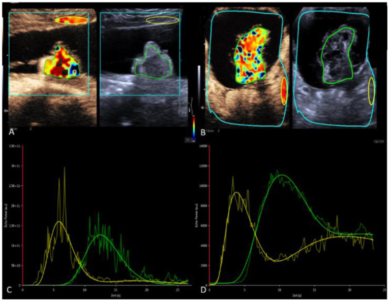
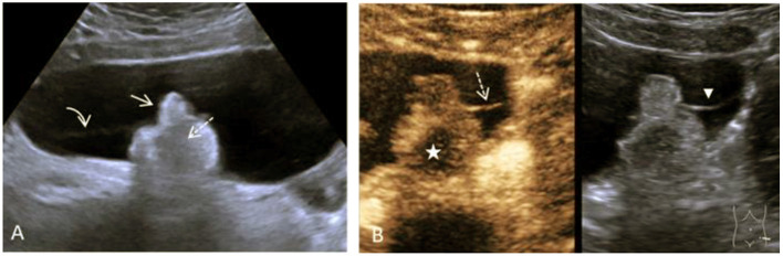
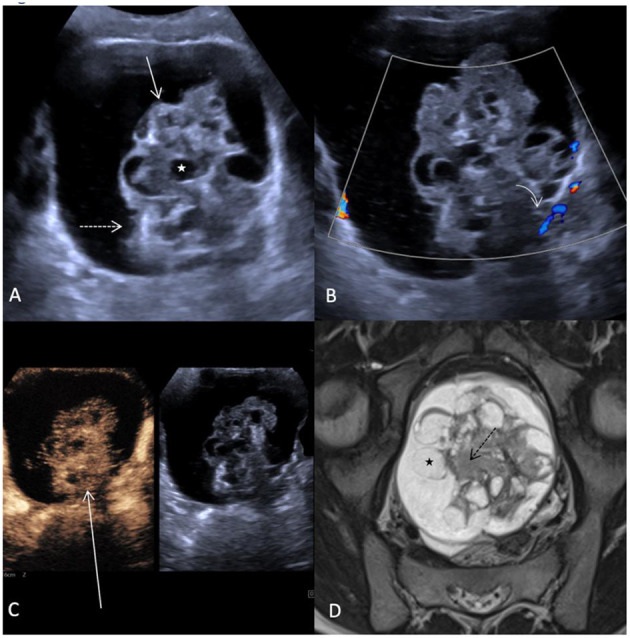
Ovarian masses are not easily differentiated on transabdominal ultrasound in children. A useful supplement in various pediatric applications is dynamic contrast-enhanced ultrasound (dynCEUS). It can be performed quickly and easily. However, the literature for dynCEUS on pediatric ovarian masses is limited. We compared two cases with ovarian teratoma in which dynCEUS was a helpful additional tool.
Keywords: dynamic CEUS; ovary; pediatric; teratoma; ultrasound.
Copyright © 2021 Glutig, Alhussami, Krüger, Waginger, Eckoldt and Mentzel.
Conflict of interest statement
The authors declare that the research was conducted in the absence of any commercial or financial relationships that could be construed as a potential conflict of interest.
Figures
Similar articles
-
Pattern recognition using transabdominal ultrasound to diagnose ovarian mature cystic teratoma.Int J Gynaecol Obstet. 2008 Nov;103(2):99-104. doi: 10.1016/j.ijgo.2008.06.002. Epub 2008 Aug 21. Int J Gynaecol Obstet. 2008. PMID: 18718589
-
Preoperative diagnosis of a "humanoid" fetus in fetu using multimode ultrasound: a case report.BMC Pediatr. 2020 Oct 19;20(1):483. doi: 10.1186/s12887-020-02389-y. BMC Pediatr. 2020. PMID: 33076884 Free PMC article.
-
Desmoplastic medulloblastoma arising from an ovarian teratoma: a case report and review of the literature.Int J Surg Pathol. 2013 Aug;21(4):427-31. doi: 10.1177/1066896912464048. Epub 2012 Nov 4. Int J Surg Pathol. 2013. PMID: 23129837 Review.
-
Retrospective evaluation of pathological results among women with ovarian endometriomas versus teratomas.Mol Clin Oncol. 2019 Jun;10(6):592-596. doi: 10.3892/mco.2019.1844. Epub 2019 Apr 15. Mol Clin Oncol. 2019. PMID: 31086669 Free PMC article.
-
Applications of contrast-enhanced ultrasound in the pediatric abdomen.Abdom Radiol (NY). 2018 Apr;43(4):948-959. doi: 10.1007/s00261-017-1315-0. Abdom Radiol (NY). 2018. PMID: 28980061 Review.
Cited by
-
[Ovarian masses in infants and children].Radiologie (Heidelb). 2024 Jan;64(1):26-34. doi: 10.1007/s00117-023-01233-5. Epub 2023 Nov 10. Radiologie (Heidelb). 2024. PMID: 37947867 Review. German.
-
Contrast-enhanced ultrasound: a comprehensive review of safety in children.Pediatr Radiol. 2021 Nov;51(12):2161-2180. doi: 10.1007/s00247-021-05223-4. Epub 2021 Oct 30. Pediatr Radiol. 2021. PMID: 34716453 Free PMC article. Review.
References
-
- Kaatsch P, Grabow D, Spix C. German Childhood Cancer Registry - Annual Report 2018 (1980-2017). Mainz: Institute of Medical Biostatistics, Epidemiology and Informatics (IMBEI) at the University Medical Center of the Johannes Gutenberg University Mainz; (2019).
Publication types
LinkOut - more resources
Full Text Sources