

Ceramide Kinase Inhibition Blocks IGF-1-Mediated Survival of Otic Neurosensory Progenitors by Impairing AKT Phosphorylation
- PMID: 34179008
- PMCID: PMC8220815
- DOI: 10.3389/fcell.2021.678760
Ceramide Kinase Inhibition Blocks IGF-1-Mediated Survival of Otic Neurosensory Progenitors by Impairing AKT Phosphorylation
Abstract
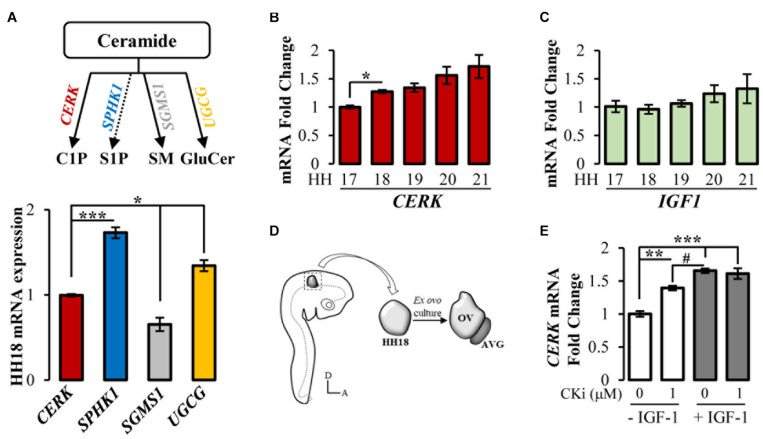
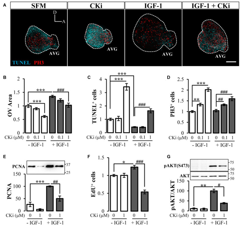
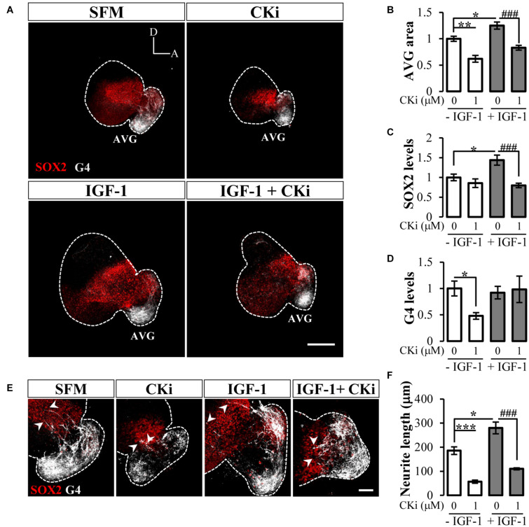
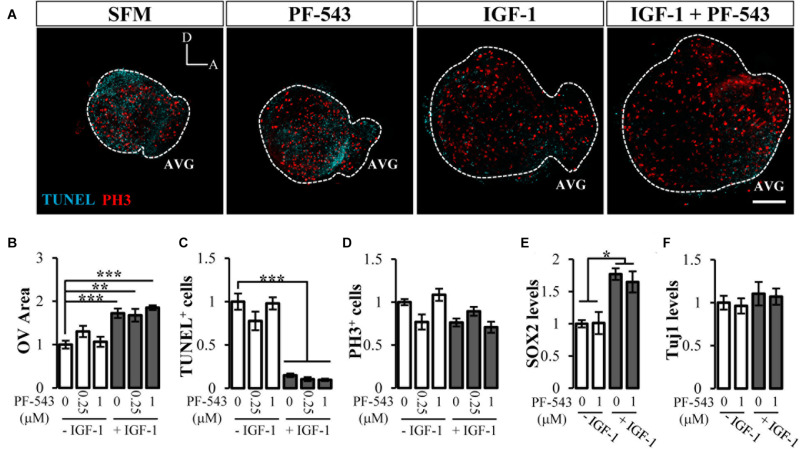
Sphingolipids are bioactive lipid components of cell membranes with important signal transduction functions in health and disease. Ceramide is the central building block for sphingolipid biosynthesis and is processed to form structurally and functionally distinct sphingolipids. Ceramide can be phosphorylated by ceramide kinase (CERK) to generate ceramide-1-phosphate, a cytoprotective signaling molecule that has been widely studied in multiple tissues and organs, including the developing otocyst. However, little is known about ceramide kinase regulation during inner ear development. Using chicken otocysts, we show that genes for CERK and other enzymes of ceramide metabolism are expressed during the early stages of inner ear development and that CERK is developmentally regulated at the otic vesicle stage. To explore its role in inner ear morphogenesis, we blocked CERK activity in organotypic cultures of otic vesicles with a specific inhibitor. Inhibition of CERK activity impaired proliferation and promoted apoptosis of epithelial otic progenitors. CERK inhibition also compromised neurogenesis of the acoustic-vestibular ganglion. Insulin-like growth factor-1 (IGF-1) is a key factor for proliferation, survival and differentiation in the chicken otocyst. CERK inhibition decreased IGF-1-induced AKT phosphorylation and blocked IGF-1-induced cell survival. Overall, our data suggest that CERK is activated as a central element in the network of anti-apoptotic pro-survival pathways elicited by IGF-1 during early inner ear development.
Keywords: NVP-231; PF-543; ceramide metabolism enzymes; development; otic progenitors; sphingolipids.
Copyright © 2021 León, Magariños and Varela-Nieto.
Conflict of interest statement
The authors declare that the research was conducted in the absence of any commercial or financial relationships that could be construed as a potential conflict of interest.
Figures
Similar articles
-
Implication of Ceramide Kinase/C1P in Cancer Development and Progression.Cancers (Basel). 2022 Jan 4;14(1):227. doi: 10.3390/cancers14010227. Cancers (Basel). 2022. PMID: 35008391 Free PMC article. Review.
-
Inhibition of Ceramide Kinase Is Effective against Cisplatin-Resistant Ovarian Cancer Cells by Regulating Ceramide and C1P Levels.Gynecol Obstet Invest. 2023;88(1):61-70. doi: 10.1159/000528869. Epub 2023 Jan 20. Gynecol Obstet Invest. 2023. PMID: 36682357
-
AKT signaling mediates IGF-I survival actions on otic neural progenitors.PLoS One. 2012;7(1):e30790. doi: 10.1371/journal.pone.0030790. Epub 2012 Jan 23. PLoS One. 2012. PMID: 22292041 Free PMC article.
-
RAF kinase activity regulates neuroepithelial cell proliferation and neuronal progenitor cell differentiation during early inner ear development.PLoS One. 2010 Dec 28;5(12):e14435. doi: 10.1371/journal.pone.0014435. PLoS One. 2010. PMID: 21203386 Free PMC article.
-
Trophic effects of insulin-like growth factor-I (IGF-I) in the inner ear.Hear Res. 2004 Oct;196(1-2):19-25. doi: 10.1016/j.heares.2003.12.022. Hear Res. 2004. PMID: 15464297 Review.
Cited by
-
Implication of Ceramide Kinase/C1P in Cancer Development and Progression.Cancers (Basel). 2022 Jan 4;14(1):227. doi: 10.3390/cancers14010227. Cancers (Basel). 2022. PMID: 35008391 Free PMC article. Review.
-
IGF-1 Controls Metabolic Homeostasis and Survival in HEI-OC1 Auditory Cells through AKT and mTOR Signaling.Antioxidants (Basel). 2023 Jan 19;12(2):233. doi: 10.3390/antiox12020233. Antioxidants (Basel). 2023. PMID: 36829792 Free PMC article.
-
Tumor Necrosis Factor-α Induces a Preeclamptic-like Phenotype in Placental Villi via Sphingosine Kinase 1 Activation.Int J Mol Sci. 2022 Mar 29;23(7):3750. doi: 10.3390/ijms23073750. Int J Mol Sci. 2022. PMID: 35409108 Free PMC article.
-
The critical roles of bioactive sphingolipids in inflammation.J Biol Chem. 2025 Jul 11;301(8):110475. doi: 10.1016/j.jbc.2025.110475. Online ahead of print. J Biol Chem. 2025. PMID: 40653199 Free PMC article. Review.
-
Ceramide-1-phosphate alleviates high-altitude pulmonary edema by stabilizing circadian ARNTL-mediated mitochondrial dynamics.J Adv Res. 2024 Jun;60:75-92. doi: 10.1016/j.jare.2023.07.008. Epub 2023 Jul 20. J Adv Res. 2024. PMID: 37479181 Free PMC article.
References
-
- Bruno M., Rizzo I. M., Romero-Guevara R., Bernacchioni C., Cencetti F., Donati C., et al. (2017). Sphingosine 1-phosphate signaling axis mediates fibroblast growth factor 2-induced proliferation and survival of murine auditory neuroblasts. Biochim. Biophys. Acta 1864 814–824. 10.1016/j.bbamcr.2017.02.004 - DOI - PubMed