. 2021 Jun 9:9:700827.
doi: 10.3389/fcell.2021.700827. eCollection 2021.

Massive Gene Loss and Function Shuffling in Appendicularians Stretch the Boundaries of Chordate Wnt Family Evolution

Affiliations

Massive Gene Loss and Function Shuffling in Appendicularians Stretch the Boundaries of Chordate Wnt Family Evolution

Josep Martí-Solans et al. Front Cell Dev Biol. .

Abstract

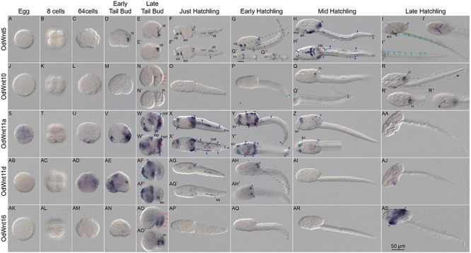
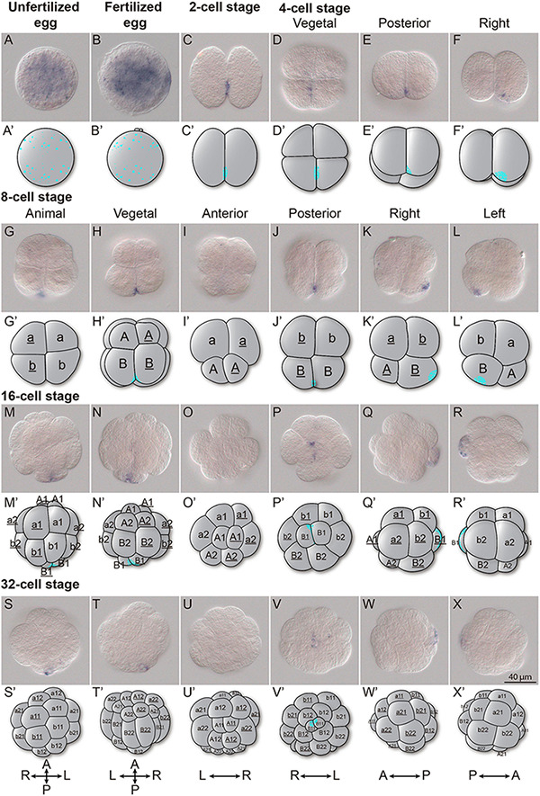
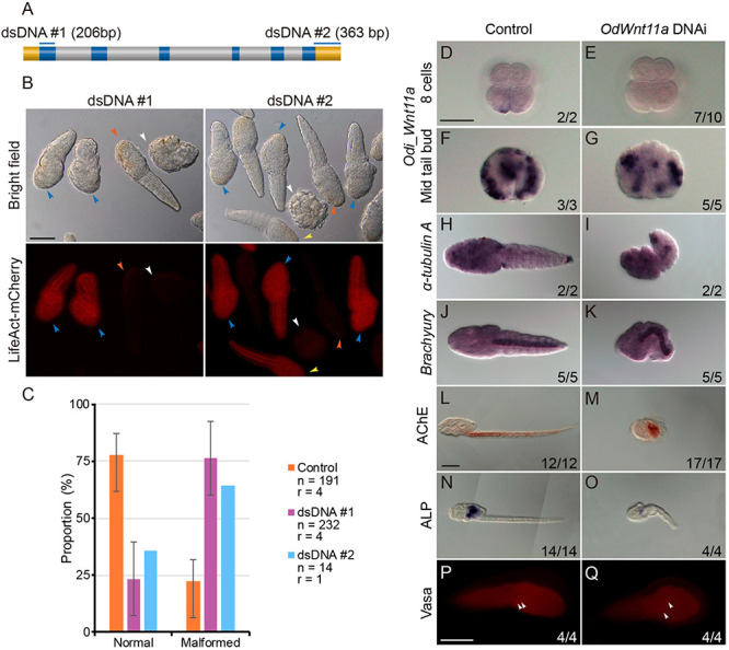
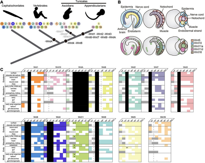
Gene loss is a pervasive source of genetic variation that influences species evolvability, biodiversity and the innovation of evolutionary adaptations. To better understand the evolutionary patterns and impact of gene loss, here we investigate as a case study the evolution of the wingless (Wnt) family in the appendicularian tunicate Oikopleura dioica, an emergent EvoDevo model characterized by its proneness to lose genes among chordates. Genome survey and phylogenetic analyses reveal that only four of the thirteen Wnt subfamilies have survived in O. dioica-Wnt5, Wnt10, Wnt11, and Wnt16,-representing the minimal Wnt repertoire described in chordates. While the loss of Wnt4 and Wnt8 likely occurred in the last common ancestor of tunicates, representing therefore a synapomorphy of this subphylum, the rest of losses occurred during the evolution of appendicularians. This work provides the first complete Wnt developmental expression atlas in a tunicate and the first insights into the evolution of Wnt developmental functions in appendicularians. Our work highlights three main evolutionary patterns of gene loss: (1) conservation of ancestral Wnt expression domains not affected by gene losses; (2) function shuffling among Wnt paralogs accompanied by gene losses; and (3) extinction of Wnt expression in certain embryonic directly correlated with gene losses. Overall our work reveals that in contrast to "conservative" pattern of evolution of cephalochordates and vertebrates, O. dioica shows an even more radical "liberal" evolutionary pattern than that described ascidian tunicates, stretching the boundaries of the malleability of Wnt family evolution in chordates.

Keywords: appendicularian tunicate chordate; chordate evolutionary developmental biology; gene function shuffling; gene loss; wingless (Wnt) family evolution.


Conflict of interest statement

The authors declare that the research was conducted in the absence of any commercial or financial relationships that could be construed as a potential conflict of interest.

Figures

FIGURE 1
FIGURE 1
Phylogenetic analysis of the Wnt family. ML phylogenetic tree of the Wnt family in chordates reveals that Wnt gene repertoire in Oikopleura dioica (names in red) belongs to four subfamilies: Wnt 5 (blue), Wnt10 (orange), Wnt11 (purple), and Wnt16 (green). The scale bar indicates amino-acid substitutions. Node support values correspond to likelihood based methods aLRT SH-like (in black) and aBayes (in magenta); support of monophyletic Wnt subfamily nodes are indicated with greater fonts size. Species abbreviations: Chordate species: Tunicates: Botryllus schlosseri (Bsc), Ciona savignyi (Csa), Ciona robusta (Cro; formerly Ciona intestinallis), Halocynthia roretzi (Hro), Halocynthia aurantium (Hau), Mogula occulta (Moccu), Mogula oculata (Mocul), Mogula occidentalis (Mocci), Phallusia fumigata (Pfu) Phallusia mammillata (Pma), and Oikopleura dioica (Odi); Cephalochordates: Branchiostoma belcheri (Bbe), Branchiostoma floridae (Bfl), Branchiostoma lanceolatum (Bla); Vertebrates: Danio rerio (Dre), Homo sapiens (Hsa). Non-chordates species: hemichordate Saccoglossus kowalevskii (Sko), annelid Capitella teleta (Cte), mollusk Lottia gigantea (Lgi), and cnidarian Nematostella vectensis (Nve).
FIGURE 2
FIGURE 2
Expression patterns of Oikopleura dioica Wnt genes during development. Whole-mount in situ hybridization in O. dioica eggs (A,J,S,AB,AK), 8 cell embryos (B,K,T,AC,AL), 64 cell embryos (C,L,U,AD,AM), early tail bud embryos (D,M,V,AE,AN), late tail bud embryos (E,N,W,A,F,AO), just hatchlings (F,O,X,AG,AP), early hatchlings (G,P,Y,AH,AQ), mid hatchlings (H,Q,Z,AI,AR) and late hatchlings (I,R,AA,AJ,AS). Upper image of each panel corresponds to left lateral view oriented anterior toward the left and dorsal toward the top. Bottom images (‘) are ventral views of optical cross sections at the levels of dashed lines. Red dots label muscle cell nuclei; magenta arrowheads label notochord; orange arrowheads label mesoendoderm; yellow arrowheads label endodermal strand; yellow asterisks label endostyle; light blue arrowheads label neuroectoderm and anterior brain; blue light asterisks label neural tube; green asterisk labels a cell posterior to the tip of the notochord that according to tubuline expression could be of neural condition. Blue dark arrowheads label sensory epidermis; blue dark asterisks label oikoplastic epithelium; blue dark double arrowheads label caudal epithelium and blue dark arrow labels posterior trunk epidermis. a, anus; cg, caudal ganglion; en, endostyle; epi, epidermis; es, endodermal strand, m, muscle cell pair; mo, mouth; not, notochord; nt, neural tube; sv, sensory vesicle; vi, vertical intestine; vo, ventral organ. Scale bar = 50.
FIGURE 3
FIGURE 3
Wnt11a expression in cleavage stages. Whole-mount in situ hybridization in O. dioica from egg up to the 32-cell stage revealing an asymmetric localization of the Wnt11a maternal transcripts. (A) Unfertilized egg. (B) Fertilized egg. (C) 2 cells stage. (D–F) 4 cells stage. (G–L) 8 cells stage. (M–R) 16 cells stage. (S–X) 32 cells stage. Schematic representations adapted form (Fujii et al., 2008) and blastomere names according to Delsman’s nomenclature are given. Embryos were viewed from various directions indicated at the top. Scale bar = 40 μm.
FIGURE 4
FIGURE 4
Wnt11a knockdown phenotypes. (A) Regions targeted by each of the PCR products. Upper box is a gene model of Wnt11a and the regions targeted by each of the PCR products are shown by blue bars above. Blue, gray and yellow boxes indicate the ORF, intron and UTR, respectively. (B) Embryos co-injection with Wnt11a PCR products (0.2 μg/μl) and Lifeact-mCherry mRNA (0.7 μg/μl). Only mCherry positive embryos were examined. Phenotypes were categorized into two groups: normal and short. Two-thirds of the animals with fluorescence show shortened trunk and tail (blue arrowheads), although some animals with fluorescence did not show malformations (yellow arrowheads). Most of the animals without fluorescence developed normally (vermillion arrowheads), while some embryos without fluorescence underwent developmental arrested before hatching (white arrowhead). (C) Proportions of each of the phenotype of hatched larvae after injection with the three kinds of PCR products. Orange, purple, and blue bars show the result of Control (Kaede PCR product), DNAi #1 and DNAi #2, respectively. Number of analyzed embryos (n) and number replicas (r) are indicated in the legend. (D–G) Whole-mount in situ hybridization for Wnt11a in 8 cells embryos (D,E) and mid tail bud stage (F,G) injected with Kaede PCR product as control and Odi_Wnt11a DNAi #1 PCR product. (H,I) Whole-mount in situ hybridization for the neuronal marker α-Tubulin A in just hatchlings injected with Kaede PCR product and Odi_Wnt11a DNAi #1 PCR product. (J,K) Whole-mount in situ hybridization for the notochord marker Brachyury in just hatchlings injected with Kaede PCR product and Odi_Wnt11a DNAi #1 PCR product. L and M. Histochemical staining for the muscle differentiation marker acetylcholinesterase (AChE) in larvae injected with Kaede PCR product and Odi_Wnt11a DNAi #1 PCR product. (N,O) Histochemical staining for the endoderm differentiation marker alkaline phosphatase (ALP) in larvae injected with Kaede PCR product and Odi_Wnt11a DNAi #1 PCR product. (P,Q) Immunohistochemistry for the germ line marker vasa in just hatchlings injected with Kaede PCR product and Odi_Wnt11a DNAi #1 PCR product. The ratio, at the bottom of each panel, indicates the proportion of embryos with the phenotype shown. Scale bar = 50 μm.
FIGURE 5
FIGURE 5
Evolutionary comparison of the Wnt gene repertoire and expression domains between Oikopleura dioica and other chordates. (A) Evolution of the Wnt subfamily repertoire (colored circles with numbers) highlighting Wnt losses (black circles) across the chordate evolutionary tree. O. dioica, with only 4 Wnt gene families—Wnt5, Wnt10, Wnt11, and Wnt16–, represents the minimal Wnt repertoire among all chordates analyzed so far. Striped circles indicate gene losses affecting only specific groups or species (e.g., Wnt1 loss in Phlebobranchia, Wnt11 in Molgulas, and Wnt3 in Botryllus). (B) Schematic illustration of Wnt expression in O. dioica. Upper panel shows drawings of O. dioica mid tailbud embryo in lateral view (left and center) and ventral view (right), with the position of central nervous system (CNS, dark blue), epidermis (light blue), endoderm (yellow), notochord (purple), and muscles (red) depicted. Lower panels show the expression of O. dioica’s Wnt genes in the mid tailbud stage. (C) Filled colored boxes denote documented expression in Somorjai et al. (2018); white filled boxes indicate absence of, or unreported expression; question marks are reserved for genes that are present, but whose expression has never been assessed; black boxes represent lost genes; dark gray boxes denote tissues/structures that are absent in the subphylum and/or at examined stages, and stippled gray boxes highlight tissues/structures lacking clear homologs, in which therefore, expression comparison is not possible. Expression patterns for vertebrate paralogs of the same Wnt subfamily were combined. O. dioica’s paralogs are indicated (a = Odi_Wnt11a; d = Odi_Wnt11d). Asterisk: caudal ganglion expression (see Supplementary Figure 4). Lan, lancelet; Asc, ascidian; Oik, O. dioica; Dan, zebrafish Danio rerio; Xen, frog Xenopus laevis/tropicalis; Gal, chicken Gallus gallus; Mus, mouse Mus musculus; PNS (ectoderm), peripheral nervous system; LPM/PSM (mesoderm), lateral plate/presomitic mesoderm; en, endostyle.

Similar articles

Cited by

References

    1. Albalat R., Cañestro C. (2016). Evolution by gene loss. Nat. Rev. Genet. 17 379–391. 10.1038/nrg.2016.39 - DOI - PubMed
    1. Andre P., Song H., Kim W., Kispert A., Yang Y. (2015). Wnt5a and Wnt11 regulate mammalian anterior-posterior axis elongation. Development 142 1516–1527. 10.1242/dev.119065 - DOI - PMC - PubMed
    1. Baranski M., Berdougo E., Sandler J. S., Darnell D. K., Burrus L. W. (2000). The dynamic expression pattern of frzb-1 suggests multiple roles in chick development. Dev. Biol. 217 25–41. 10.1006/dbio.1999.9516 - DOI - PubMed
    1. Bassham S., Postlethwait J. (2000). Brachyury (T) expression in embryos of a larvacean urochordate, Oikopleura dioica, and the ancestral role of T. Dev. Biol. 220 322–332. 10.1006/dbio.2000.9647 - DOI - PubMed
    1. Cañestro C., Postlethwait J. H. (2007). Development of a chordate anterior-posterior axis without classical retinoic acid signaling. Dev. Biol. 305 522–538. 10.1016/j.ydbio.2007.02.032 - DOI - PubMed

LinkOut - more resources