. 2021 Jun 4:8:644797.
doi: 10.3389/fcvm.2021.644797. eCollection 2021.

α-Ketoglutarate Upregulates Collecting Duct (Pro)renin Receptor Expression, Tubular Angiotensin II Formation, and Na+ Reabsorption During High Glucose Conditions

Affiliations

α-Ketoglutarate Upregulates Collecting Duct (Pro)renin Receptor Expression, Tubular Angiotensin II Formation, and Na+ Reabsorption During High Glucose Conditions

Aarón Guerrero et al. Front Cardiovasc Med. .

Abstract

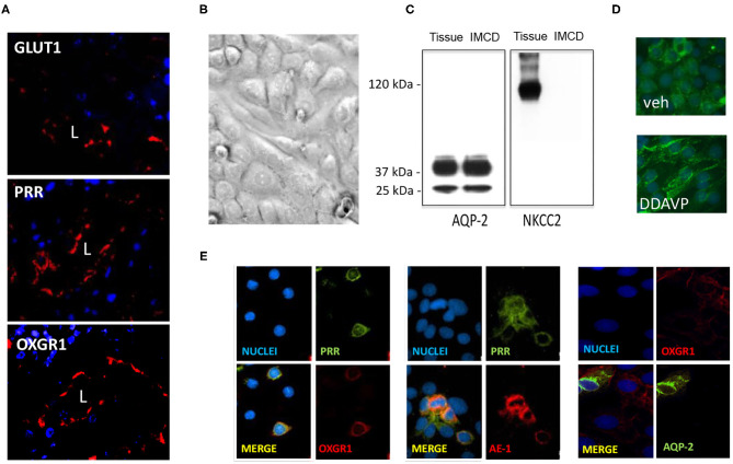
Diabetes mellitus (DM) causes high glucose (HG) levels in the plasma and urine. The (pro)renin receptor (PRR) is a key regulator of renal Na+ handling. PRR is expressed in intercalated (IC) cells of the collecting duct (CD) and binds renin to promote angiotensin (Ang) II formation, thereby contributing to Na+ reabsorption. In DM, the Kreb's cycle is in a state of suppression in most tissues. However, in the CD, expression of glucose transporters is augmented, boosting the Kreb's cycle and consequently causing α-ketoglutarate (αKG) accumulation. The αKG receptor 1 (OXGR1) is a Gq-coupled receptor expressed on the apical membrane of IC cells of the CD. We hypothesize that HG causes αKG secretion and activation of OXGR1, which increases PRR expression in CD cells. This effect then promotes intratubular AngII formation and Na+ reabsorption. To test this hypothesis, streptozotocin (STZ)-induced diabetic mice were treated with or without montelukast (ML), an OXGR1 antagonist, for 6 days. STZ mice had higher urinary αKG and PRR expression along with augmented urinary AngII levels and Na+ retention. Treatment with ML prevented all these effects. Similarly, primary cultured inner medullary CD cells treated with HG showed increased PRR expression, while OXGR1 antagonist prevented this effect. αKG increases PRR expression, while treatments with ML, PKC inhibition, or intracellular Ca2+ depletion impair this effect. In silico analysis suggested that αKG binds to mouse OXGR1. These results indicate that HG conditions promote increased levels of intratubular αKG and OXGR1-dependent PRR upregulation, which impact AngII formation and Na+ reabsorption.

Keywords: Kreb's cycle; angiotensin; collecting duct; diabetes; prorenin receptor.


Conflict of interest statement

The authors declare that the research was conducted in the absence of any commercial or financial relationships that could be construed as a potential conflict of interest.

Figures

Figure 1
Figure 1
Physiological parameters in control mice and streptozotocin-induced diabetic mice with or without treatment with montelukast (ML), an OXGR1 antagonist. (A) Body weight was reduced after 6 days of streptozotocin (STZ) treatment. (B) Fasting glucose levels were augmented on days 3 and 6 in STZ and STZ + ML mice. (C) After 6 days, a significant increase in urinary levels of αKG (assessed by αKG/creatinine ratio) was detected in STZ and STZ + ML mice. (D) A slight but not significant reduction was observed in food intake. (E) Water intake was augmented in STZ and STZ + ML groups. (F) Urine output was also augmented in STZ and STZ + ML group. (G) Systolic, diastolic, and mean arterial blood pressure was not changed at the end of the treatment. *p < 0.05 vs. control group (n = 7–9).
Figure 2
Figure 2
(A) After 6 days of a single streptozotocin (STZ) administration, (pro)renin receptor (PRR) messenger RNA (mRNA) levels were augmented in the renal medulla of STZ mice. (B) Same effect was observed in PRR protein abundance. Treatment with OXGR antagonist montelukast (ML) in STZ mice prevented the upregulation of PRR mRNA and protein. (C) GLUT1 mRNA and (D) protein levels were increased in the renal medulla of STZ mice, as well as in STZ and STZ + ML groups. ML alone did not show significant changes on PRR or GLUT expression. *p < 0.05 vs. control group (n = 6).
Figure 3
Figure 3
Streptozotocin (STZ) mice showed augmented urinary AngII excretion and Na+ retention that was prevented by OXGR1 pharmacological blockade. (A) AngII was significantly augmented in STZ mice (p < 0.001 vs. control group). Although mice treated with the OXGR1 antagonist montelukast (ML) also showed significant increases in AngII levels, this effect was less evident (p < 0.05). Treatment with ML alone in normoglycemic mice did not significantly change AngII excretion. (B) No changes were observed in plasma AngII. (C) Na+ balance assessed on day 6 demonstrated that STZ mice retained Na+. ML treatment in STZ mice blunted this effect. Saline challenge was performed to determine the percentage of injected Na+ that would be excreted. (D) Urine samples collected every hour showed a continuous decrease in total volume with no significant differences between controls and STZ or STZ + ML treated mice. (E) Na+ concentration of these samples was measured in order to calculate the percentage of Na+ excreted after saline (0.9% NaCl) injection. STZ mice showed a reduced percentage of injected Na+ in urine samples, demonstrating increased Na+ retention. This effect was less evident in STZ + ML group. Significant values were observed only at 2, 3, and 4 h as compared to controls. *p < 0.05 vs. control group, ***p < 0.001 vs. control group, n = 9.
Figure 4
Figure 4
(A) Staining of whole kidney sections showed the presence of GLUT1, (pro)renin receptor (PRR), and OXGR1 (red staining). L indicates lumen. Nuclei are stained with 4,6-diamidino-2-phenylindole dihydrochloride (DAPI). (B) Inner medullary collecting duct (IMCD) cells were grown until reach confluence and assessed for the presence of NKCC to rule out the presence of cortical tissues cells. Western blot analysis showed the presence of aquaporin (AQP)-2 in homogenates of total kidney tissue and IMCD cells. (C) NKCC was absent in IMCD. Functional IMCD cells were assessed by the activation of the vasopressin V2 with a V2 receptor agonist (DDAVP, 10−8 M). (D) After 45 min, AQP-2 immunofluorescence was mostly detected in plasma membrane. Coimmunostaining demonstrated the coexistence of OXGR1 and PRR, which also co-localizes with AE-1, a marker of intercalated cell. (E) OXGR1 did not co-localizes with AQP-2, a marker of principal cell.
Figure 5
Figure 5
Evidence of the effect of αKG on (pro)renin receptor (PRR) expression and intracellular signaling pathways. (A) Incubations with HG during 24 h increased in αKG accumulation in inner medullary collecting duct (IMCD) cell culture media. HG + ML did not cause αKG accumulation (*p < 0.05 vs. control group, n = 5). (B) Functional assessment of cultured IMCD cells demonstrated that αKG induced a rise in intracellular Ca2+ that was blunted by OXGR1 antagonist montelukast (ML). This suggest that the mechanism involves the activation of OXGR1. (C) IMCD incubated with αKG 0.2 mM for 16 h showed increased levels of PRR; this effect was blunted by ML. (D) Increased PRR levels was suppressed by PKC inhibitor calphostin C (10 nM) or intracellular Ca2+ depletion using thapsigargin (1 μM). (E) OXGR1 expression did not change in HG conditions. For Panels (C–E); *p < 0.05 vs. control group, n = 3.
Figure 6
Figure 6
In-silico evidence of the binding of αKG to OXGR1 (GPR99). (A) Three-dimensional structure for mouse GPR99. (B) Using the server Swiss-Dock, the model in Panel (A) was used for docking studies of binding of αKG to OXGR1. (C) A large pocket inside the OXGR1 (blue color) can be observed in which ARG110, HIS114, ARG228, and ARG261 are the most important residues in the interaction with αKG. (D) Multiple alignment analysis in rat, mice, and human GPR99 (OXGR1) and GPR91 (P2Y) demonstrated that residues R99, H103, R252, and R281 of GPR91 are the same residues present in the hydrophobic pocket of OXGR1.
Figure 7
Figure 7
Effect of high glucose (HG) conditions (25 mM) on GLUT1 and (pro)renin receptor (PRR) expression in cultured inner medullary collecting duct (IMCD) cells subjected or not to OXGR1 pharmacological blockade. (A) PRR messenger RNA (mRNA) and (B) protein levels were augmented by 24-h HG treatment while pretreatment with 10−7 M of montelukast (ML) blunted the induction of PRR expression. Augmentation of GLUT1 at (C) mRNA and (D) protein levels was not altered by the pretreatment with ML. *p < 0.05 vs. control group, n = 5. Reblotting of blot (B) is shown in Panel (D), and same representative β-actin loading control was used.
Figure 8
Figure 8
Working hypothesis representing the effects of high glucose on the activation of tricarboxylic acid (TCA) cycle and accumulation of αKG leading to OXGR1 activation. Elevated plasma glucose levels and increased expression of GLUT1 boost glycolysis and TCA cycle, causing αKG accumulation and secretion. Prorenin (and renin) released into the lumen by the principal cell, binds to (pro)renin receptor (PRR), consequently increasing renin activity and activating enzymatic activity of prorenin. Angiotensinogen in the urine (secreted by proximal tubules or coming from blood filtrate) is cleaved by activated prorenin or renin. Since ACE activity is present along the nephron, this intratubular RAS activation ends with AngII intratubular formation and angiotensin type 1 receptor (ATR1)-dependent stimulation of sodium reabsorption through epithelial sodium channels (ENaC).

References

    1. Zimpelmann J, Kumar D, Levine DZ, Wehbi G, Imig JD, Navar, et al. . Early diabetes mellitus stimulates proximal tubule renin mRNA expression in the rat. Kidney Int. (2000) 58:2320–30. 10.1046/J.1523-1755.2000.00416.X - DOI - PubMed
    1. Kang JJ, Toma I, Sipos A, Meer EJ, Vargas Peti-Peterdi SL. The collecting duct is the major source of prorenin in diabetes. Hypertension. (2008) 51:1597–604. 10.1161/Hypertensionaha.107.107268 - DOI - PMC - PubMed
    1. Persson Lu F X, Rossing P, Garrelds IM, Danser AHJ, Parving H-H. Urinary renin and angiotensinogen in type 2 diabetes: added value beyond urinary albumin? J Hypertens. (2013) 31:1646–52. 10.1097/HJH.0b013e328362217c - DOI - PubMed
    1. Mamenko M, Zaika O, Ilatovskaya DV, Staruschenko Pochynyuk AO. Angiotensin II increases activity of the epithelial Na+ channel (ENaC) in distal nephron additively to aldosterone. J Biol Chem. (2012) 287:660–71. 10.1074/jbc.M111.298919 - DOI - PMC - PubMed
    1. Mamenko M, Zaika O, Prieto MC, Jensen VB, Doris PA, Navar, et al. . Chronic Angiotensin II infusion drives extensive aldosterone-independent epithelial Na+ channel activation. Hypertension. (2013) 62:1111–22. 10.1161/Hypertensionaha.113.01797 - DOI - PMC - PubMed