Conducting polymer-based granular hydrogels for injectable 3D cell scaffolds
- PMID: 34179344
- PMCID: PMC8225239
- DOI: 10.1002/admt.202100162
Conducting polymer-based granular hydrogels for injectable 3D cell scaffolds
Abstract
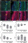
Injectable 3D cell scaffolds possessing both electrical conductivity and native tissue-level softness would provide a platform to leverage electric fields to manipulate stem cell behavior. Granular hydrogels, which combine jamming-induced elasticity with repeatable injectability, are versatile materials to easily encapsulate cells to form injectable 3D niches. In this work, we demonstrate that electrically conductive granular hydrogels can be fabricated via a simple method involving fragmentation of a bulk hydrogel made from the conducting polymer PEDOT:PSS. These granular conductors exhibit excellent shear-thinning and self-healing behavior, as well as record-high electrical conductivity for an injectable 3D scaffold material (~10 S m-1). Their granular microstructure also enables them to easily encapsulate induced pluripotent stem cell (iPSC)-derived neural progenitor cells, which were viable for at least 5 days within the injectable gel matrices. Finally, we demonstrate gel biocompatibility with minimal observed inflammatory response when injected into a rodent brain.
Keywords: 3D cell scaffolds; conductive hydrogels; injectable hydrogels.
Conflict of interest statement
Conflict of Interest The authors report no competing interests.
Figures


Similar articles
-
Conductive and Adhesive Granular Alginate Hydrogels for On-Tissue Writable Bioelectronics.Gels. 2023 Feb 19;9(2):167. doi: 10.3390/gels9020167. Gels. 2023. PMID: 36826337 Free PMC article.
-
Electrically Conductive Injectable Silk/PEDOT: PSS Hydrogel for Enhanced Neural Network Formation.J Biomed Mater Res A. 2025 Jan;113(1):e37859. doi: 10.1002/jbm.a.37859. J Biomed Mater Res A. 2025. PMID: 39719872
-
Injectable and Conductive Granular Hydrogels for 3D Printing and Electroactive Tissue Support.Adv Sci (Weinh). 2019 Aug 21;6(20):1901229. doi: 10.1002/advs.201901229. eCollection 2019 Oct 16. Adv Sci (Weinh). 2019. PMID: 31637164 Free PMC article.
-
Granular hydrogels: emergent properties of jammed hydrogel microparticles and their applications in tissue repair and regeneration.Curr Opin Biotechnol. 2019 Dec;60:1-8. doi: 10.1016/j.copbio.2018.11.001. Epub 2018 Nov 24. Curr Opin Biotechnol. 2019. PMID: 30481603 Free PMC article. Review.
-
Rational design of injectable conducting polymer-based hydrogels for tissue engineering.Acta Biomater. 2022 Feb;139:4-21. doi: 10.1016/j.actbio.2021.04.027. Epub 2021 Apr 22. Acta Biomater. 2022. PMID: 33894350 Review.
Cited by
-
Conductive Microgel Annealed Scaffolds Enhance Myogenic Potential of Myoblastic Cells.Adv Healthc Mater. 2024 Oct;13(25):e2302500. doi: 10.1002/adhm.202302500. Epub 2023 Dec 18. Adv Healthc Mater. 2024. PMID: 38069833 Free PMC article.
-
Hydrogel Bioelectronics for Health Monitoring.Biosensors (Basel). 2023 Aug 14;13(8):815. doi: 10.3390/bios13080815. Biosensors (Basel). 2023. PMID: 37622901 Free PMC article. Review.
-
Smart bioelectronics and biomedical devices.Biodes Manuf. 2022;5(1):1-5. doi: 10.1007/s42242-021-00179-8. Epub 2022 Jan 14. Biodes Manuf. 2022. PMID: 35043079 Free PMC article. No abstract available.
-
Conducting Polymer Nanoparticles with Intrinsic Aqueous Dispersibility for Conductive Hydrogels.Adv Mater. 2024 Jan;36(1):e2306691. doi: 10.1002/adma.202306691. Epub 2023 Nov 23. Adv Mater. 2024. PMID: 37680065 Free PMC article.
-
Sticking Together: Injectable Granular Hydrogels with Increased Functionality via Dynamic Covalent Inter-Particle Crosslinking.Small. 2022 Sep;18(36):e2201115. doi: 10.1002/smll.202201115. Epub 2022 Mar 22. Small. 2022. PMID: 35315233 Free PMC article.
References
-
- da Silva LP, Kundu SC, Reis RL, Correlo VM, Trends Biotechnol. 2020, 38, 24. - PubMed
-
- Burnstine-Townley A, Eshel Y, Amdursky N, Adv. Funct. Mater 2019, 30, 1901369.
-
- Gajendiran M, Choi J, Kim SJ, Kim K, Shin H, Koo HJ, Kim K, J. Ind. Eng. Chem 2017, 51, 12.
Grants and funding
LinkOut - more resources
Full Text Sources
