

Mediastinal mixed germ cell tumor: A case report and literature review
- PMID: 34179505
- PMCID: PMC8216226
- DOI: 10.1515/med-2021-0293
Mediastinal mixed germ cell tumor: A case report and literature review
Abstract
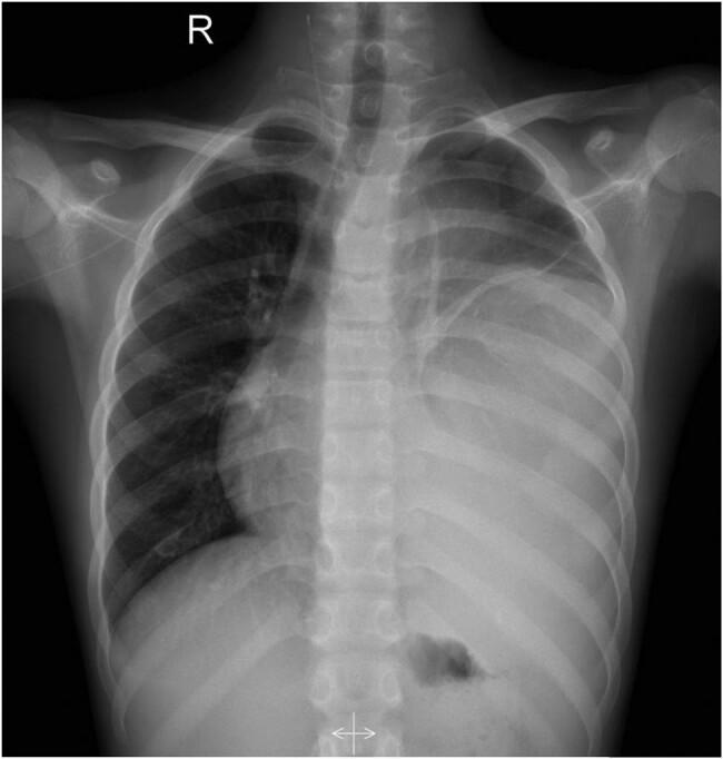
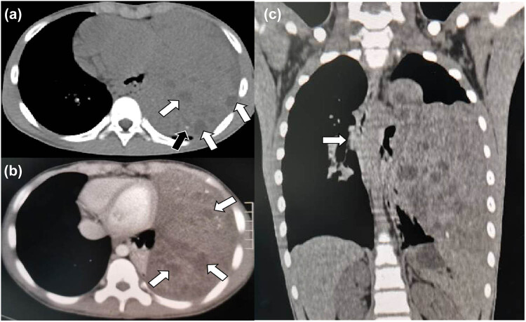
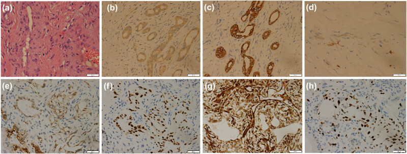
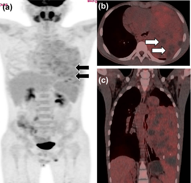
Mixed germ cell tumor (MGCT) mainly occurs in young women's ovaries and men's testicles and rarely occurs outside the gonad. Fewer than 10 cases of mediastinal MGCT are available in PubMed, Embase, and other databases in English, while mediastinal MGCT with three pathological components, such as yolk sac tumor, immature teratoma, and embryonal carcinoma, has not been reported previously. A 12-year-old male sought medical attention for chest discomfort and underwent a computed tomography (CT) scan. A large soft tissue mass occupying most of the left thoracic cavity and mediastinum was detected. A CT-guided biopsy was performed, and an MGCT was diagnosed with pathological components, including yolk sac tumor, immature teratoma, and a small amount of embryonal carcinoma. Due to the large size of the tumor, the patient was treated with an EP regimen (etoposide + cisplatin) and paclitaxel + ifosfamide + cisplatin interstitial chemotherapy. The patient was followed up for 6 months and was alive with the disease. To the best of our knowledge, this is the 10th patient with MGCT in the mediastinum. The incidence of mediastinal MGCT is low, but it should still be considered one of the differential diagnoses of isolated pleural fibroma and neurogenic tumors.
Keywords: case report; computed tomography; mediastinal; mixed germ cell tumor.
© 2021 Xianwen Hu et al., published by De Gruyter.
Conflict of interest statement
Conflict of interest: The authors declare no conflict of interest.
Figures

References
-
- Ghaemmaghami F , Ayatollahi H , Daneshbodi B , Azmoodeh FA . Unusual location of ovarian mixed germ cell tumor. Int J Gynecol Cancer. 2005;15(5):979–83. - PubMed
- Ghaemmaghami F, Ayatollahi H, Daneshbodi B, Azmoodeh FA. Unusual location of ovarian mixed germ cell tumor. Int J Gynecol Cancer. 2005;15(5):979–83. - PubMed
-
- Kurman RJ , Ellenson LH , Ronnett BM . Blaustein’s pathology of the female genital. Vol. 20(3). 6th edition. New York: Springer Science + business Media, LLC; 2011. p. 848.
- Kurman RJ, Ellenson LH, Ronnett BM. Blaustein’s pathology of the female genital. 3. 6th edition. Vol. 20. New York: Springer Science + business Media, LLC; 2011. p. p. 848.
-
- Pradhan D , Kaman L , Dhillon J , Mohanty SK . Mediastinal mixed germ cell tumor in an infertile male with Klinefelter syndrome: a case report and literature review. J Cancer Res Ther. 2015;11(4):1034. - PubMed
- Pradhan D, Kaman L, Dhillon J, Mohanty SK. Mediastinal mixed germ cell tumor in an infertile male with Klinefelter syndrome: a case report and literature review. J Cancer Res Ther. 2015;11(4):1034. - PubMed
-
- Yuri T , Shimano N , Ohashi Y , Miki K , Tsukamoto R , Tsubura A . An autopsy case of primary mixed choriocarcinoma and mature teratoma located in the thymic region associated with elevated human chorionic gonadotropin levels and characteristic testicular changes. Med Mol Morphol. 2006;39(1):49–53. - PubMed
- Yuri T, Shimano N, Ohashi Y, Miki K, Tsukamoto R, Tsubura A. An autopsy case of primary mixed choriocarcinoma and mature teratoma located in the thymic region associated with elevated human chorionic gonadotropin levels and characteristic testicular changes. Med Mol Morphol. 2006;39(1):49–53. - PubMed
-
- Taccagni GL , Parafioriti A , Dell’Antonio G , Crespi G . Mixed germ cell tumour of the mediastinum (seminoma, embryonal carcinoma, choriocarcinoma and teratoma). Light and electron microscopic cytology and histological investigation. Pathol Res Pract. 1989;185(4):506–10. - PubMed
- Taccagni GL, Parafioriti A, Dell’Antonio G, Crespi G. Mixed germ cell tumour of the mediastinum (seminoma, embryonal carcinoma, choriocarcinoma and teratoma). Light and electron microscopic cytology and histological investigation. Pathol Res Pract. 1989;185(4):506–10. - PubMed
Publication types
LinkOut - more resources
Full Text Sources