

Endothelial corneal cell damage after pars plana vitrectomy: analogy of different intraocular tamponade agents
- PMID: 34179579
- PMCID: PMC8207861
- DOI: 10.22336/rjo.2021.29
Endothelial corneal cell damage after pars plana vitrectomy: analogy of different intraocular tamponade agents
Abstract
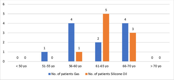
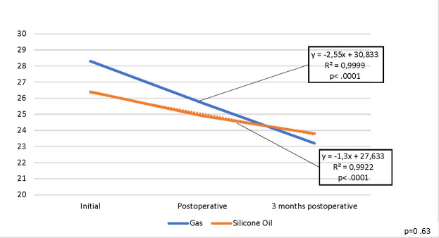
A reduction in the corneal endothelial cells multitude after anterior pole intervention is well established in numerous researches, but there are few articles that follow the impact of vitreoretinal interventions on the cornea, especially when endotamponade agents are used. The assessment of the endothelial corneal cells is needed since it facilitates the personal evaluation of the functional endothelial stock. Specular microscopy investigation offers a scale of the functional strength of the endothelium of cornea, which is vital before all intraocular interventions. Endotamponade agents are very suitable and important tools in the surgical treatment of retinal detachment, but their use must be differentiated depending on the uniqueness of each patient. This research shows corneal endothelial damages caused by intraocular tamponade agents of different types in the case of pars plana vitrectomy for cases of multitude retinal detachments. The purpose of the research was to determine the changes that appear in the endothelium of the cornea and to deal with the results when different tamponade agents are used in the surgical cure for retinal detachment. Specular endothelial corneal microscopy records were achieved and the modifications of the following parameters revealed corneal implication: mean endothelial cell densities, average cell area, coefficient of variation, hexagonality and corneal center thickness. On the first day and at three months postoperatively, a statistically significant reduction was observed for the CV, MCD, and HEX parameters (p 0.001), but no statistically significant difference of the two endotamponade agents (for MCD, p=0.15; for CV, p=0.63; for HEX, p=0.93) was noticed. AVG parameter had a statistically significant decrease (p 0.001) and there was also a statistically significant difference of the two endotamponade agents (p=0.03), patients with gas tamponade presenting a superior result. On the first day and at three months postoperatively, the corneal center thickness presented a statistically significant increase (p 0.001) and there was a statistically significant difference between the two endotamponade agents (p=0.03), patients with gas endotamponade presenting a superior result. In conclusion, using the intraocular tamponade agents helps reestablish the functional-anatomical recovery of the retina after surgery, but their special indication must be well-established for each case of retinal detachment.
Keywords: corneal specular microscopy; endotamponade agents; number of endothelial corneal cells; retinal detachment; silicone oil.
© The Authors.Romanian Society of Ophthalmology.
Figures


References
-
- Norton EWD. Intraocular gas in the management of selected detachments. Ophthalmology. 1973;77:OP85. - PubMed
-
- Haut J, Ullern M, Boulard ML. Utilisation du silicone intra-oculaire après vitrectomie comme traitement des rétractions massives du vitré. Bull Soc Ophthalmol Fr. 1978;28:361. - PubMed
-
- Machemer R, Buettner H, Norton EDW, Parel JM. Vitrectomy: a pars plana approach. Trans Am Acad Ophthalmol Otolaryngol. 1971;75:813–820. - PubMed
-
- Feibel RM, Blodi CF, Cibis PA. MD: a pioneer of modern vitreoretinal surgery. JAMA Ophthalmol. 2013 Aug;131(8):1077–1082. doi: 10.1001/jamaophthalmol.2013.597. - PubMed
-
- Mentes R, Stellmans P. Comparison of post-operative comfort in 20 gauge Versus 23 Gauge pars plana vitrectomy. Bull Soc Belge Ophthalmol. 2009;311:5–10. - PubMed
MeSH terms
Substances
LinkOut - more resources
Full Text Sources
Medical
Miscellaneous