

Generation and expression analysis of BAC humanized mice carrying HLA-DP401 haplotype
- PMID: 34179719
- PMCID: PMC8212823
- DOI: 10.1002/ame2.12158
Generation and expression analysis of BAC humanized mice carrying HLA-DP401 haplotype
Abstract
Background: Human leukocyte antigen (HLA)-DP is much less studied than other HLA class II antigens, that is, HLA-DR and HLA-DQ, etc. However, the accumulating data have suggested the important roles of DP-restricted responses in the context of cancer, allergy, and infectious disease. Lack of animal models expressing these genes as authentic cis-haplotypes blocks our understanding for the role of HLA-DP haplotypes in immunity.
Methods: To explore the potential cis-acting control elements involved in the transcriptional regulation of the HLA-DPA1/DPB1 gene, we performed the expression analysis using bacterial artificial chromosome (BAC)-based transgenic humanized mice in the C57BL/6 background, which carried the entire HLA-DP401 gene locus. We further developed a mouse model of Staphylococcus aureus pneumonia in HLA-DP401 humanized transgenic mice, and performed the analysis on the expression pattern of HLA-DP401 and immunological responses in the model.
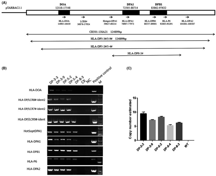
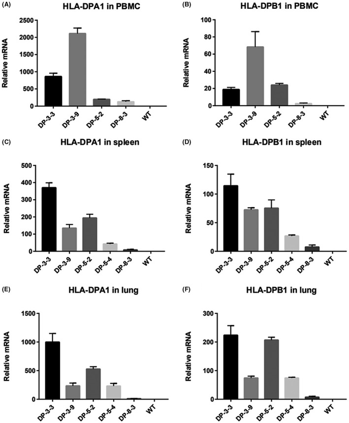
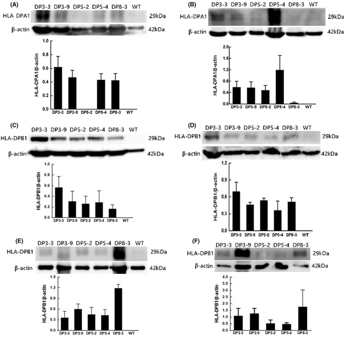
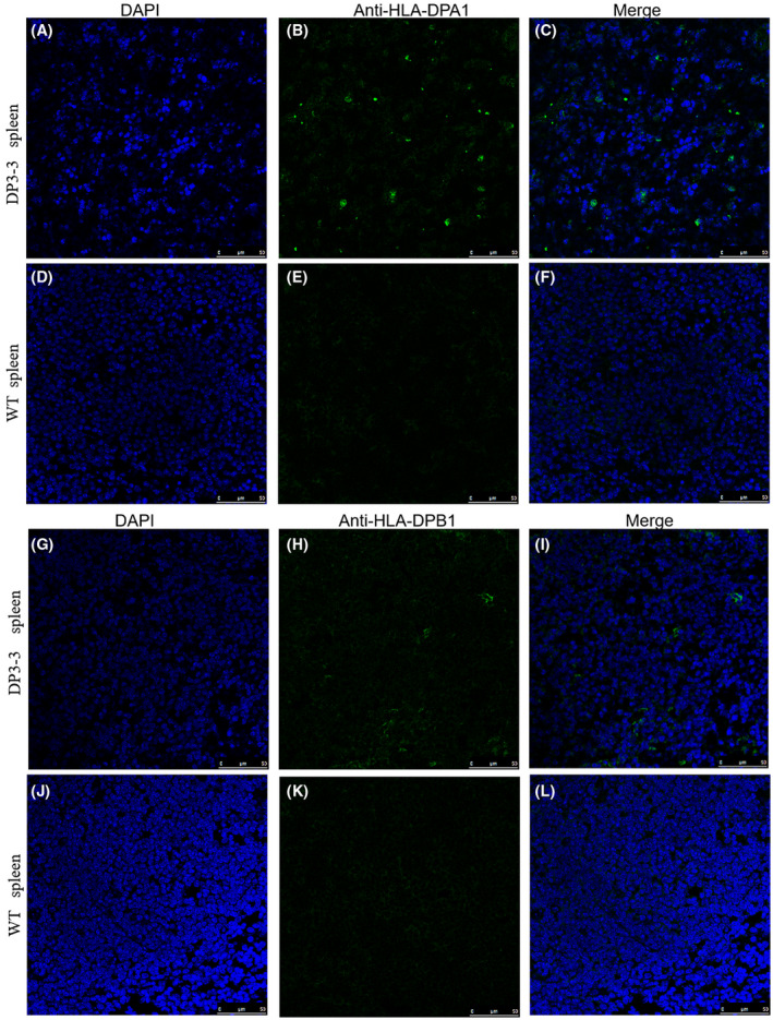
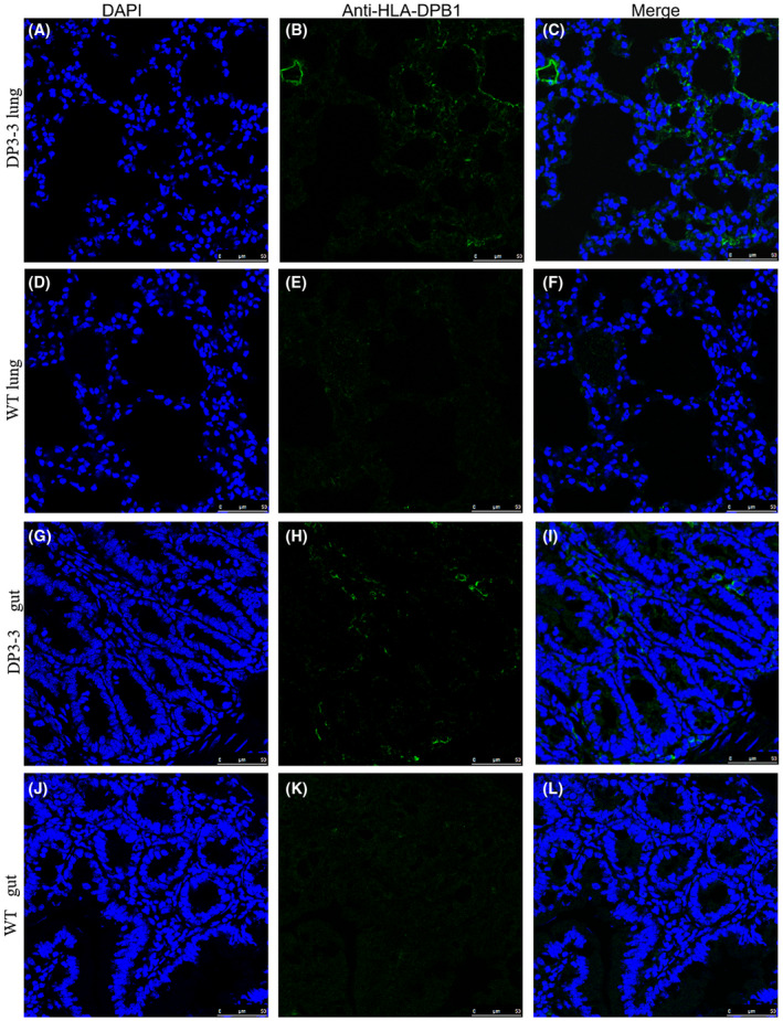
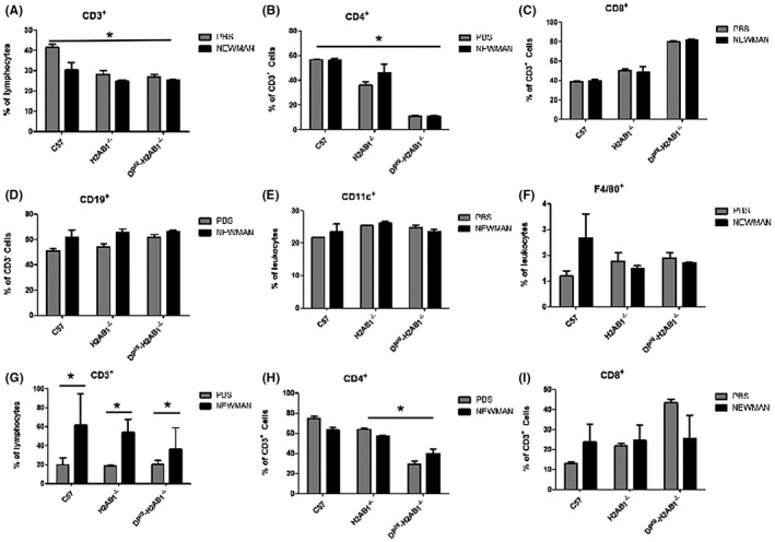
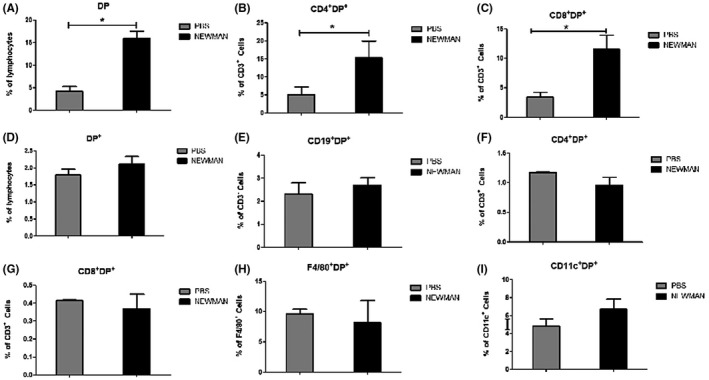
Results: In this study, we screened and identified a BAC clone spanning the entire HLA-DP gene locus. DNA from this clone was analyzed for integrity by pulsed-field gel electrophoresis and then microinjected into fertilized mouse oocytes to produce transgenic founder animals. Nine sets of PCR primers for regional markers with an average distance of 15 kb between each primer were used to confirm the integrity of the transgene in the five transgenic lines carrying the HLA-DPA1/DPB1 gene. Transgene copy numbers were determined by real-time PCR analysis. HLA-DP401 gene expression was analyzed at the mRNA and protein level. Although infection with S aureus Newman did not alter the percentage of immune cells in the spleen and thymus from the HLA-DP401-H2-Aβ1 humanized mice. Increased expression of HLA-DP401 was observed in the thymus of the humanized mice infected by S aureus.
Conclusions: We generated several BAC transgenic mice, and analyzed the expression of HLA-DPA1/DPB1 in those mice. A model of Saureus-induced pneumonia in the HLA-DP401-H2-Aβ1-/- humanized mice was further developed, and S aureus infection upregulated the HLA-DP401 expression in thymus of those humanized mice. These findings demonstrate the potential of those HLA-DPA1/DPB1 transgenic humanized mice for developing animal models of infectious diseases and MHC-associated immunological diseases.
Keywords: HLA‐DP4; Staphylococcus aureus pneumonia; bacterial artificial chromosome (BAC); gene expression; humanized mice.
© 2021 The Authors. Animal Models and Experimental Medicine published by John Wiley & Sons Australia, Ltd on behalf of The Chinese Association for Laboratory Animal Sciences.
Figures
Similar articles
-
Characterization of genetic humanized mice with transgenic HLA DP401 or DRA but deficient in endogenous murine MHC class II genes upon Staphylococcus aureus pneumonia.Animal Model Exp Med. 2023 Dec;6(6):585-597. doi: 10.1002/ame2.12331. Epub 2023 May 29. Animal Model Exp Med. 2023. PMID: 37246733 Free PMC article.
-
A 320-kilobase artificial chromosome encoding the human HLA DR3-DQ2 MHC haplotype confers HLA restriction in transgenic mice.J Immunol. 2002 Mar 15;168(6):3050-6. doi: 10.4049/jimmunol.168.6.3050. J Immunol. 2002. PMID: 11884478
-
HLA-DP on Epithelial Cells Enables Tissue Damage by NKp44+ Natural Killer Cells in Ulcerative Colitis.Gastroenterology. 2023 Oct;165(4):946-962.e13. doi: 10.1053/j.gastro.2023.06.034. Epub 2023 Jul 15. Gastroenterology. 2023. PMID: 37454979 Free PMC article.
-
Role of HLA class II genes in susceptibility/resistance to inflammatory arthritis: studies with humanized mice.Immunol Rev. 2010 Jan;233(1):62-78. doi: 10.1111/j.0105-2896.2009.00858.x. Immunol Rev. 2010. PMID: 20192993 Review.
-
Advances in bacterial artificial chromosome (BAC) transgenic mice for gene analysis and disease research.Gene. 2025 Jan 20;934:149014. doi: 10.1016/j.gene.2024.149014. Epub 2024 Oct 24. Gene. 2025. PMID: 39461574 Review.
Cited by
-
Simultaneous loading of PCR-based multiple fragments on mouse artificial chromosome vectors in DT40 cell for gene delivery.Sci Rep. 2022 Dec 16;12(1):21790. doi: 10.1038/s41598-022-25959-9. Sci Rep. 2022. PMID: 36526651 Free PMC article.
-
Genetic mutation of Tas2r104/Tas2r105/Tas2r114 cluster leads to a loss of taste perception to denatonium benzoate and cucurbitacin B.Animal Model Exp Med. 2024 Jun;7(3):324-336. doi: 10.1002/ame2.12357. Epub 2023 Dec 28. Animal Model Exp Med. 2024. PMID: 38155461 Free PMC article.
-
Development of a humanized HLA-A30 transgenic mouse model.Animal Model Exp Med. 2022 Dec;5(4):350-361. doi: 10.1002/ame2.12225. Epub 2022 Jul 6. Animal Model Exp Med. 2022. PMID: 35791899 Free PMC article.
-
Progress, implications, and challenges in using humanized immune system mice in CAR-T therapy-Application evaluation and improvement.Animal Model Exp Med. 2024 Feb;7(1):3-11. doi: 10.1002/ame2.12353. Epub 2023 Oct 12. Animal Model Exp Med. 2024. PMID: 37823214 Free PMC article. Review.
-
Generation of transchromosomic mice harboring HLA-A/B/C and human B2M via mouse artificial chromosome and triple BAC integration.Sci Rep. 2025 Jul 30;15(1):27852. doi: 10.1038/s41598-025-13138-5. Sci Rep. 2025. PMID: 40739281 Free PMC article.
References
-
- Mangalam AK, Rajagopalan G, Taneja V, David CS. HLA class II transgenic mice mimic human inflammatory diseases. Adv Immunol. 2008;97:65‐147. - PubMed
Publication types
MeSH terms
Substances
LinkOut - more resources
Full Text Sources
Research Materials
