

doi: 10.1016/j.jaccas.2021.05.003.
Epub 2021 Jun 16.
Multiple Etiologies to Myocardial Injury in COVID-19
Affiliations
- PMID: 34179834
- PMCID: PMC8208893
- DOI: 10.1016/j.jaccas.2021.05.003
Item in Clipboard
Multiple Etiologies to Myocardial Injury in COVID-19
JACC Case Rep.
2021 Jun.
Abstract
In patients with acute myocardial injury secondary to coronavirus disease-2019 (COVID-19), cardiovascular magnetic resonance imaging can identify the underlying pathology. We highlight a case of acute myocardial injury secondary to COVID-19, which demonstrated both epicardial vessel thrombosis and the recently described phenomenon of microvascular thrombosis. (Level of Difficulty: Advanced.).
Keywords: CMR, cardiac magnetic resonance imaging; COVID-19, coronavirus-2019; cardiac magnetic resonance; myocardial infarction; thrombosis.
© 2021 The Authors.
Conflict of interest statement
The authors have reported that they have no relationships relevant to the contents of this paper to disclose.
Figures

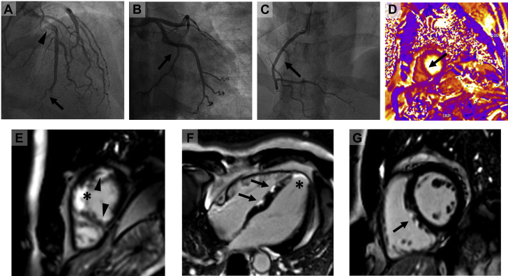
Coronary Angiography and Cardiac MRI Images in COVID-19 (A) Distal occlusion of the left anterior descending coronary artery detected on coronary angiography (arrow) and fresh thrombus midvessel (arrowhead). (B) Normal left circumflex artery (arrow) and (C) normal right coronary artery (arrow). (D) T2 map at apical level shows edema in the septum (arrow). (E) Short-axis cardiac magnetic resonance imaging (CMR) late gadolinium enhancement image shows infarction in the apical septum (asterisk) as well as ischemic pattern late gadolinium enhancement (LGE) at the inferoseptal junction and the apical lateral wall (arrowheads). (F) 4-chamber CMR LGE image shows infarction in the apical cap (asterisk) and patchy subepicardial enhancement in the interventricular septum (arrows). (G) Short-axis CMR LGE images show patchy sub-epicardial enhancement in the interventricular septum (arrow).
References
-
- Dario P., Rika K., Giulio G. Microthrombi as a major cause of cardiac injury in COVID-19: a pathologic study. Circulation. 2021;143:1031–1042. - PubMed
Grants and funding
LinkOut - more resources
Full Text Sources
