. 2021 May 16:10:225-235.
doi: 10.1016/j.ibneur.2021.05.002. eCollection 2021 Jun.

Comparison of effect of crush or transection peripheral nerve lesion on lumbar spinal cord synaptic plasticity and microglial dynamics

Affiliations

Comparison of effect of crush or transection peripheral nerve lesion on lumbar spinal cord synaptic plasticity and microglial dynamics

Raquel M P Campos et al. IBRO Neurosci Rep. .

Abstract

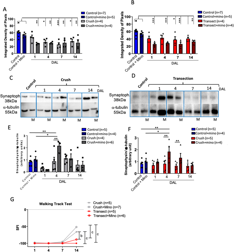
In an injury to the peripheral nervous system, the spinal cord and brain structure reorganize connections to optimize the function of the remaining parts. Many cell events are triggered in the spinal cord to support changes in the synaptic connections around motoneurons, where old connections are removed, and new ones created. Microglial cells are primitive macrophages that invade the central nervous system in early stages of neurodevelopment and have several functions, such as eliminating synapses. We investigated the synaptic plasticity after different types of peripheral (sciatic) nerve injury (crush or total transection), as well as the behavior of microglial cells for 2 weeks after a peripheral lesion. As expected, sciatic-nerve injury reduced motor performance in mice, but crushed animals regained partial motor control. Because of sciatic-nerve injury, pre-synaptic inputs decreased around the motoneurons in the ventro-lateral horn, while microglial cells increased around these cells. Microglial cells also exhibited altered morphology in both types of peripheral lesion, indicating a similar underlying mechanism of plasticity. To investigate the involvement of microglia in this scenario, microglial activation was modulated by daily administration of minocycline. The minocycline treatment directly affected the microglial response and impacted the synapse rearrangement in the spinal cord. Together, these results demonstrate that microglia cells are involved in synaptic plasticity in the lumbar spinal cord in both nerve-injury scenarios.

Summary of statement: Here, we demonstrated that acute plasticity in the lumbar spinal cord (LSC) did not differ between crush and transection of peripheral nerve, and that microglial reactivity in the LSC was important after both injury types.

Keywords: Lumbar spinal cord; Microglia; Sciatic nerve; Synaptic plasticity.


Figures

Fig. 1
Fig. 1
Experimental design. (A) Timeline showing days following nerve injury in mice, where the walking-track test and the time of euthanasia are indicated. (B) Timeline showing the experimental design of the minocycline treatment, where the first dose was administered 6 h prior to the surgical procedure, followed by a dose every 24 h after the injury.
Fig. 2
Fig. 2
Synaptophysin-staining decreases around motoneurons after sciatic injury. Images taken from LSC transverse sections (ipsilateral to the nerve injury) in the ventral horn immunolabeled for synaptophysin (white) and nuclei counterstained with DAPI (blue). Animals with crushed nerves (C–F) and transected nerves (G–J) 1, 4, 7, and 14 days after lesion (DAL). Image of ventral horn from uninjured animal (control) at medium (A) and low (B) magnification (contour of spinal cord white matter indicated by dotted yellow line). Soma of motoneurons indicated by yellow arrows. Quantitative analysis of integrated density of pixels surrounding motoneurons in all experimental conditions and DAL (K). Graph of sciatic function index (SFI) analysis obtained by walking-track test (L). Statistics: Two-way ANOVA, Tukey’s post-test. *p < 0.05 and **p < 0.005. For synaptophysin analysis, Control n = 7, Crush n = 4/DAL (total 16), Transection n = 4/DAL (total 16). For walking track, Control n = 4, Crush n = 5/DAL (total 20), Transection n = 5/DAL (total 20). Scale bars: A, C–J = 50 µm; B = 100 µm.
Fig. 3
Fig. 3
Microglia recruitment in dorsal and ventral spinal cord horns after nerve injury. Images obtained by apotome microscopy of transverse sections from LSC ipsilateral to nerve injury, immunolabeled for Iba-1 (red) and counterstained with DAPI (white) in dorsal and ventral horns from crush (A–D, I–L) and transected (E–H, M–P) groups 1, 4, 7, and 14 days after lesion (DAL). Low magnification (20×) of LSC ipsilateral to lesion stained for Iba-1 (red) and DAPI (blue) (Q). Histogram of quantitative analysis comparing microglia density in all LSC ipsilateral to nerve injury (Iba-1+) (R). Control n = 4, Crush n = 4/DAL (total 16), Transection n = 4/DAL (total 16). Statistics: Two-way ANOVA, **p < 0.005 and ***p < 0.001. Scale bars: A–P = 50 µm; Q = 100 µm.
Fig. 4
Fig. 4
Microglia recruitment in acute periods after nerve injury is reversed by minocycline in both injury types. Quantitative analysis of Iba-1-positive cells by images taken with apotome microscopy of LSC ipsilateral to nerve injury following minocycline treatment in Crushed (A) and Transected (B) animals. Images taken by apotome at high magnification of LSC transverse sections in ventral horn, of Iba-1-positive microglial cells (red) of uninjured (Control, C), Crush (D), Transection (E), Crush + minocycline (F), and Transection + minocycline (G) animals 7DAL. Nuclei in white were counterstained with DAPI (white). 3D branching reconstruction using ImageJ, from uninjured (Control, H), Crush (I), Transection (J), Crush plus minocycline (K), and Transection plus minocycline (L) animals. Histograms of quantitative analysis comparing the number of microglial branches (M,N). For microglia quantification: Control n = 4, Control+Mino n = 4; Crush n = 4/DAL (total 16), Crush+Mino n = 4/DAL (total 16); Transection n = 4/DAL (total 16), Transection+Mino n = 4/DAL (total 16). For morphological analysis: Control n = 4, Control+Mino n = 4; Crush n = 4, Crush+Mino n = 4; Transection n = 4, Transection+Mino n = 4. Statistics: Two-way ANOVA, *p < 0.05 and **p < 0.005 for graphs A and B; One-way ANOVA, *p < 0.05 for graphs M and N. Scale bars: C–G = 10 µm.
Fig. 5
Fig. 5
Minocycline treatment alters synaptophysin expression in LSC after crush and transection sciatic-nerve lesion. Histograms of quantitative analysis of synaptophysin in an integrated density of pixels surrounding motoneurons from groups treated or not with minocycline (A–B). Immunoblots for synaptophysin (38kD) and alpha tubulin (42kD) from LSC tissue dissected from L4–L6 region. Membrane with LSC samples of unlesioned, after nerve crush, and nerve crush + minocycline groups (C) 1, 4, 7, and 14 days after lesion. Membrane with LSC samples of unlesioned, after nerve transection, and nerve transection + minocycline groups (D) 1, 4, 7, and 14 days after lesion. M indicates minocycline treatment in C and D. Histograms of quantitative analysis of synaptophysin:alpha-tubulin density ratio measured by immunoblots, represented in arbitrary units for crushed nerves (E), transected nerves (F), and treated or not with minocycline. Graph of sciatic-function index (SFI) scores of injured animals treated or not with minocycline (G). For synaptophysin immunofluorescence analysis, Control n = 7, Control+Mino n = 5; Crush n = 4/DAL (total 16), Crush+Mino n = 4/DAL (total 16); Transection n = 4/DAL (total 16), Transection+Mino n = 4/DAL (total 16). For synaptophysin western-blotting analysis, Control n = 5, Control+Mino n = 4; Crush n = 4/DAL (total 16), Crush+Mino n = 6/DAL (total 24); Transection n = 5/DAL (total 20), Transection+Mino n = 4/DAL (total 16). For walking track, Crush n = 5/DAL (total 20), Crush+Mino n = 7/DAL (total 21); Transection n = 5/DAL (total 20), Transection+Mino n = 6/DAL (total 24). Statistics: Two-way ANOVA, *p < 0.05, **p < 0.005, ***p < 0.001.

Similar articles

Cited by

References

    1. Ahmed A., Wang L.L., Abdelmaksoud S., Aboelgheit A., Saeed S., Zhang C.L. Minocycline modulates microglia polarization in ischemia-reperfusion model of retinal degeneration and induces neuroprotection. Sci. Rep. 2017;7(1):14065. - PMC - PubMed
    1. Aldskogius H. Mechanisms and consequences of microglial responses to peripheral axotomy. Front. Biosci. 2011;3:857–868. - PubMed
    1. Aldskogius H., Kozlova E.N. Central neuron-glial and glial-glial interactions following axon injury. Prog. Neurobiol. 1998;55(1):1–26. - PubMed
    1. Alvarez F.J., Titus-Mitchell H.E., Bullinger K.L., Kraszpulski M., Nardelli P., Cope T.C. Permanent central synaptic disconnection of proprioceptors after nerve injury and regeneration. I. Loss of VGLUT1/IA synapses on motoneurons. J. Neurophysiol. 2011;106(5):2450–2470. - PMC - PubMed
    1. Arbat-Plana A., Torres-Espín A., Navarro X., Udina E. Activity dependent therapies modulate the spinal changes that motoneurons suffer after a peripheral nerve injury. Exp. Neurol. 2015;263:293–305. - PubMed