

Intracranial hemorrhage detected through a craniotomy site with point of care ultrasound
- PMID: 34179872
- PMCID: PMC8212560
- DOI: 10.1002/emp2.12419
Intracranial hemorrhage detected through a craniotomy site with point of care ultrasound
Abstract
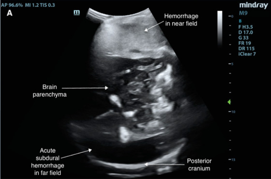
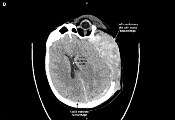
A 60-year-old male presented to the emergency department with acute change in mental status while recovering from a recent hemicraniectomy. During evaluation by the emergency physician, a point-of-care ultrasound (POCUS) was performed using the patient's existing craniectomy site as a sonographic window. Multiple areas of intracranial hemorrhage were visualized on POCUS and head computed tomography scan ultimately requiring urgent neurosurgical intervention. Our case report demonstrates an innovative application of POCUS in the emergency department- setting that has potential to expedite diagnosis and management of life-threatening neurosurgical etiologies, such as hemorrhage and midline shift, in a unique patient population.
Keywords: POCUS; TCUS; brain; craniotomy; emergency; intracranial hemorrhage; neurosurgery; point of care ultasound; transcranial ultrasound; ultasound.
© 2021 The Authors. JACEP Open published by Wiley Periodicals LLC on behalf of American College of Emergency Physicians.
Conflict of interest statement
Authors have no conflicts of interest to disclose.
Figures
Similar articles
-
Identification of intracranial hemorrhage progression by transcranial point-of-care ultrasound in a patient with prior hemicraniectomy: a case report.J Ultrasound. 2022 Jun;25(2):399-402. doi: 10.1007/s40477-021-00588-6. Epub 2021 Apr 28. J Ultrasound. 2022. PMID: 33913120 Free PMC article.
-
Ethical and medicolegal aspects in the management of neurosurgical emergencies among Jehovah's Witnesses: Clinical implications and review.Clin Neurol Neurosurg. 2020 Jul;194:105798. doi: 10.1016/j.clineuro.2020.105798. Epub 2020 Mar 19. Clin Neurol Neurosurg. 2020. PMID: 32222653 Review.
-
Brain Point of Care Ultrasound in Young Children Receiving Computed Tomography in the Emergency Department: A Proof of Concept Study.POCUS J. 2023 Nov 27;8(2):165-169. doi: 10.24908/pocus.v8i2.16435. eCollection 2023. POCUS J. 2023. PMID: 38099165 Free PMC article.
-
Hemorrhagic Cholecystitis: A Case of Expedited Diagnosis by Point-of-Care Ultrasound in the Emergency Department.J Emerg Med. 2019 Jul;57(1):74-76. doi: 10.1016/j.jemermed.2019.03.010. Epub 2019 Apr 15. J Emerg Med. 2019. PMID: 31000429
-
Arterial Gas Emboli Secondary to Portal Venous Gas Diagnosed With Point-of-Care Ultrasound: Case Report and Literature Review.J Emerg Med. 2020 Dec;59(6):906-910. doi: 10.1016/j.jemermed.2020.06.060. Epub 2020 Aug 5. J Emerg Med. 2020. PMID: 32771317 Review.
References
-
- Nambiar M, MacIsaac C, Grabinski R, Liew D, Kavar B. Outcomes of decompressive craniectomy in patients after traumatic brain injury. Crit Care Resusc. 2015;17(2):67‐72. - PubMed
-
- Saqqur M, Zygun D, Demchuk A. Role of transcranial doppler in neurocritical care. Crit Care Med. 2007;35(5 Suppl):S216‐S223. - PubMed
-
- Caricato A, Mignani V, Sandroni C, Pietrini D. Bedside detection of acute epidural hematoma by transcranial sonography in a head‐injured patient. Intensive Care Med. 2010;36(6):1091‐1092. - PubMed
Publication types
LinkOut - more resources
Full Text Sources