. 2021 Sep;156(3):197-207.
doi: 10.1007/s00418-021-02008-6. Epub 2021 Jun 27.

Reprogramming of DNA methylation is linked to successful human preimplantation development

Affiliations

Reprogramming of DNA methylation is linked to successful human preimplantation development

Julia Arand et al. Histochem Cell Biol. 2021 Sep.

Abstract

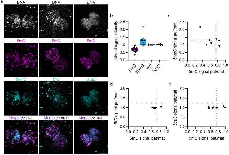
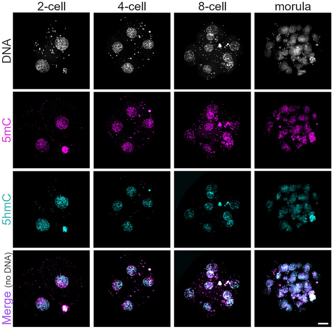
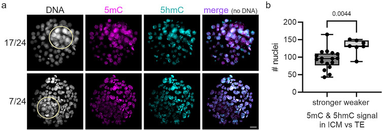
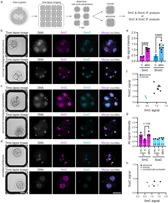
Human preimplantation development is characterized by low developmental rates that are poorly understood. Early mammalian embryogenesis is characterized by a major phase of epigenetic reprogramming, which involves global DNA methylation changes and activity of TET enzymes; the importance of DNA methylation reprogramming for successful human preimplantation development has not been investigated. Here, we analyzed early human embryos for dynamic changes in 5-methylcytosine and its oxidized derivatives generated by TET enzymes. We observed that 5-methylcytosine and 5-hydroxymethylcytosine show similar, albeit less pronounced, asymmetry between the parental pronuclei of human zygotes relative to mouse zygotes. Notably, we detected low levels of 5-formylcytosine and 5-carboxylcytosine, with no apparent difference in maternal or paternal pronuclei of human zygotes. Analysis of later human preimplantation stages revealed a mosaic pattern of DNA 5C modifications similar to those of the mouse and other mammals. Strikingly, using noninvasive time-lapse imaging and well-defined cell cycle parameters, we analyzed normally and abnormally developing human four-cell embryos for global reprogramming of DNA methylation and detected lower 5-methylcytosine and 5-hydroxymethylcytosine levels in normal embryos compared to abnormal embryos. In conclusion, our results suggest that DNA methylation reprogramming is conserved in humans, with human-specific dynamics and extent. Furthermore, abnormalities in the four-cell-specific DNA methylome in early human embryogenesis are associated with abnormal development, highlighting an essential role of epigenetic reprogramming for successful human embryogenesis. Further research should identify the underlying genomic regions and cause of abnormal DNA methylation reprogramming in early human embryos.

Keywords: Active DNA demethylation; DNA methylation reprogramming; Human preimplantation development; Successful human embryogenesis.


Conflict of interest statement

The authors declare no competing financial interests.

Figures

Fig. 1
Fig. 1
Human-specific dynamics of DNA methylation reprogramming in zygotes. a Representative images of indirect immunostaining for 5mC and 5hmC, 5fC, or 5caC in human zygotes at late G2 stage (a total of 5–20 zygotes per DNA modification were analyzed. Scale bar = 20 μm). b Quantification of integral IF signal intensities of 5mC (magenta) and derivatives (cyan) in human zygotes at late G2 stage normalized against DNA signal. Shown are paternal versus maternal signal ratios for 5mC, 5hmC, 5fC, and 5caC. Note the high variability in paternal/maternal 5mC and 5hmC signal ratios (each dot in the box plot represents a single embryo). ce Scatter plots of 5mC versus 5hmC (c), 5fC (d), and 5caC signals (e). The dotted line on the x-axis at 0.75 marks a cutoff for loss of 5mC and the dotted line on the y-axis at 1.25 a cutoff for gain of 5hmC/5fC/5caC in the paternal pronucleus
Fig. 2
Fig. 2
Immunofluorescence analysis of 5mC and 5hmC in human cleavage-stage embryos. Representative immunofluorescence images of human embryos at distinct stages of preimplantation development analyzed for 5mC (magenta) and 5hmC signals (cyan) (n = 9, 24, 9, 11 for two-cell, four-cell, eight-cell, and morula-stage embryos, respectively). Scale bar = 20 μm
Fig. 3
Fig. 3
Immunofluorescence analysis of human blastocysts. a Representative immunofluorescence images of human blastocysts analyzed for 5mC (magenta), 5hmC(cyan), and DNA signals (white). The major fraction of human blastocysts (17 of 24, upper panel) show stronger 5mC and 5hmC signals in the inner cell mass (ICM) compared to the trophectoderm; a smaller subset (7 of 24, lower panel) show weaker 5mC and 5hmC signals in the ICM compared to trophectoderm. The ICM is indicated by a yellow circle. Scale bar = 20 μm. b Number of cells per blastocyst with stronger or weaker 5mC and 5hmC signals in the ICM compared to trophectoderm. Each dot represents a single blastocyst. Statistically significant differences were calculated using two-tailed Student’s t test
Fig. 4
Fig. 4
DNA methylation reprogramming in normally and abnormally developing human four-cell embryos. a Experimental setup. bd Representative immunostaining of human embryos cultured to the four-cell stage by noninvasive time-lapse imaging and analyzed by 5mC, 5hmC, and 5caC IF. b Human four-cell embryos predicted to develop to the blastocyst stage via noninvasive time-lapse parameters (see Wong et al. 2010). c Human embryos cultured to the four-cell stage and predicted to be nonviable via noninvasive time-lapse parameters and d arrested human embryos. Scale bar = 20 μm. eh Quantification of 5mC (e, g), 5hmC (e), and 5caC (f) signal intensities in normally (n.) and abnormally (abn.) developing four-cell embryos. Antibody signals were normalized to DNA signal. Each dot represents a single embryo. Statistical significance was calculated using the Mann–Whitney test (e) or Kruskal–Wallis test (g). Correlation of 5mC to 5hmC (f) and 5mC to 5caC (h) signal intensity in analyzed four-cell embryos. Each dot represents a single embryo; normally developing embryos are shown in blue. m. = multi-nucleated

References

    1. Amouroux R, Nashun B, Shirane K, Nakagawa S, Hill PW, D'Souza Z, Nakayama M, Matsuda M, Turp A, Ndjetehe E, Encheva V, Kudo NR, Koseki H, Sasaki H, Hajkova P. De novo DNA methylation drives 5hmC accumulation in mouse zygotes. Nat Cell Biol. 2016;18:225–233. doi: 10.1038/ncb3296. - DOI - PMC - PubMed
    1. Aoki F, Worrad DM, Schultz RM. Regulation of transcriptional activity during the first and second cell cycles in the preimplantation mouse embryo. Dev Biol. 1997;181:296–307. doi: 10.1006/dbio.1996.8466. - DOI - PubMed
    1. Arand J, Wossidlo M, Lepikhov K, Peat JR, Reik W, Walter J. Selective impairment of methylation maintenance is the major cause of DNA methylation reprogramming in the early embryo. Epigenetics Chromatin. 2015;8:1. doi: 10.1186/1756-8935-8-1. - DOI - PMC - PubMed
    1. Cassuto NG, Montjean D, Siffroi JP, Bouret D, Marzouk F, Copin H, Benkhalifa M. Different levels of DNA methylation detected in human sperms after morphological selection using high magnification microscopy. Biomed Res Int. 2016;2016:6372171. doi: 10.1155/2016/6372171. - DOI - PMC - PubMed
    1. Chavez SL, Loewke KE, Han J, Moussavi F, Colls P, Munne S, Behr B, Reijo Pera RA. Dynamic blastomere behaviour reflects human embryo ploidy by the four-cell stage. Nat Commun. 2012;3:1251. doi: 10.1038/ncomms2249. - DOI - PMC - PubMed

Substances

LinkOut - more resources