. 2021 Sep;44(9):1448-1455.
doi: 10.1007/s00270-021-02859-5. Epub 2021 Jun 27.

Endovascular Stent-Graft Repair of the Ascending Aorta: Assessment of a Specific Novel Stent-Graft Design in Phantom, Cadaveric, and Clinical Application

Affiliations

Endovascular Stent-Graft Repair of the Ascending Aorta: Assessment of a Specific Novel Stent-Graft Design in Phantom, Cadaveric, and Clinical Application

Sven R Hauck et al. Cardiovasc Intervent Radiol. 2021 Sep.

Abstract

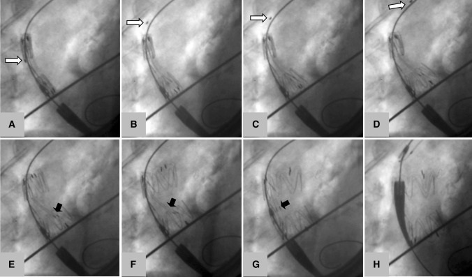
Purpose: To test a stent-graft specifically designed for the ascending aorta in phantom, cadaver, and clinical application, and to measure deployment accuracy to overcome limitations of existing devices.

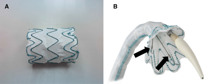
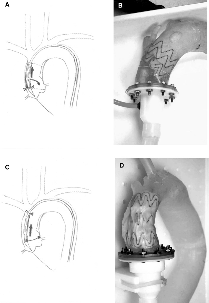
Methods: A stent-graft has been designed with support wires to fixate the apices toward the inner curvature, thereby eliminating the forward movement of the proximal end which can happen with circumferential tip capture systems. The device was deployed in three aortic phantoms, and in four cadavers, deployment precision was measured. Subsequently, the device was implanted in a patient to exclude a pseudoaneurysm originating from the distal anastomosis after ascending aortic replacement.

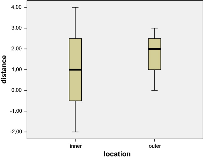
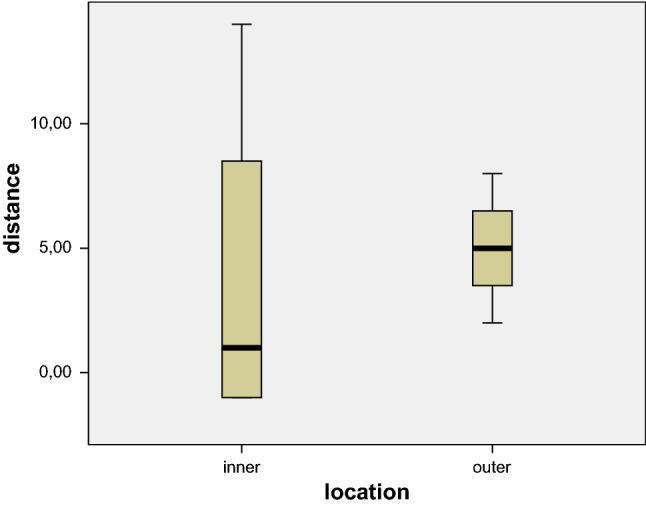
Results: The stent-grafts were successfully deployed in all phantoms and cadavers. Deployment accuracy of the proximal end of the stent-graft was within 1 mm proximally and 14 mm distally to the intended landing zone on the inner curvature, and 2-8 mm distal to the intended landing zone on the outer curvature. In clinical application, the pseudoaneurysm could be successfully excluded without complications.

Conclusion: The novel stent-graft design promises accurate placement in the ascending aorta. The differential deployment of the apices at the inner and outer curvatures allows deployment perpendicular to the aortic axis.

Level of evidence: No level of evidence.

Keywords: Ascending TEVAR; Ascending aortic stent-graft; Custom-made stentgraft.


Conflict of interest statement

The authors declare that they have no conflict of interest.

Figures

Fig. 1
Fig. 1
The dedicated ascending aortic stent-graft in fully (A) and partially (B) deployed state. The support wires for the apices facing the inner curvature are shown in B (black arrows). Images courtesy of Terumo Aortic
Fig. 2
Fig. 2
During ascending aortic stenting from a femoral approach, the stentgraft is closely appositioned to the outer curvature of the aorta. In conventional stent-grafts (A, B), the proximal apices are clasped to a proximal pivotal point at the base of the nosecone (arrowheads). During deployment, when the sheath is retracted (straight arrow), the parts of the stentgraft facing the inner curvature must move in a quarter-circle toward the inner aortic wall, since they are still attached to the nosecone (curved arrow). This creates an oblique position of the stentgraft with the inner parts dangerously close to the origin of the left coronay artery (A, dotted line, B-phantom experiment). The novel design (C, D) supports two apices facing the inner curvature with long levers from distal (arrowheads), instead of clasping them from proximal. During retraction of the sheath (straight arrow), the inner parts of the stentgraft are guided in a slightly curved path (curved arrow) toward the inner wall. This results in a predictable positioning of the proximal end of the stentgraft almost exactly perpendicular to the longitudinal axis of the ascending aorta (D-phantom experiment).
Fig. 3
Fig. 3
Boxplots indicating the actual location of the proximal end of the stent-graft in the phantom experiments in relation to the intended landing zone on the inner (left) and the outer (right) curvature of the aorta. The whiskers represent the actual range, the solid boxes the 2nd and 3rd quartiles, respectively
Fig. 4
Fig. 4
Boxplots indicating the actual location of the proximal end of the stent-graft in the cadaver experiments in relation to the intended landing zone on the inner (left) and the outer (right) curvature of the aorta. The whiskers represent the actual range, the solid boxes the 2nd and 3rd quartiles, respectively
Fig. 5
Fig. 5
Deployment of the novel stent-graft in the ascending aorta of a human cadaver. (A) Stent-graft inside the soft 12-mm nylon sheath in deployment position (B) opening of the stent-graft by retraction of the soft sheath. Note the marker on the soft sheath gradually retracted in A–D (white arrows) (C, D). The part of the stent-graft facing the inner curvature moves controlled and perpendicular to the aortic axis toward the inner curvature of the ascending aorta (E). Note the two levers aiding in stabilizing the stent-graft during this maneuver (black arrows). (F) The stent-graft has made contact with the inner aortic curvature, the inner apices of the proximal stent segment are still held by the two levers. (G) The tip capture has been released; the two levers are now parallel to the central cannula. (H) The stent-graft is fully released; the delivery system is retracted
Fig. 6
Fig. 6
CT and digital subtraction angiography before (A, B) and after (C, D) exclusion of an ascending aortic pseudoaneurysm by deployment of 2 dedicated ascending aortic stent-grafts

References

    1. Schoder M, Lammer J, Czerny M. Endovascular aortic arch repair: hopes and certainties. Eur J Vasc Endovasc Surg. 2009;38(3):255–261. doi: 10.1016/j.ejvs.2009.06.007. - DOI - PubMed
    1. Czerny M, Zimpfer D, Rodler S, Funovics M, Dorfmeister M, Schoder M, Marta G, Weigang E, Gottardi R, Lammer J, Wolner E, Grimm M. Endovascular stent-graft placement of aneurysms involving the descending aorta originating from chronic type B dissections. Ann Thorac Surg. 2007;83(5):1635–1639. doi: 10.1016/j.athoracsur.2006.12.041. - DOI - PubMed
    1. Schoder M, Czerny M, Cejna M, Rand T, Stadler A, Sodeck GH, Gottardi R, Loewe C, Lammer J. Endovascular repair of acute type B aortic dissection: long-term follow-up of true and false lumen diameter changes. Ann Thorac Surg. 2007;83(3):1059–1066. doi: 10.1016/j.athoracsur.2006.10.064. - DOI - PubMed
    1. Gottardi R, Zimpfer D, Funovics M, Schoder M, Lammer J, Wolner E, Czerny M, Grimm M. Mid-term results after endovascular stent-graft placement due to penetrating atherosclerotic ulcers of the thoracic aorta. Eur J Cardiothorac Surg. 2008;33(6):1019–1024. doi: 10.1016/j.ejcts.2007.12.054. - DOI - PubMed
    1. Canaud L, Joyeux F, Berthet JP, Hireche K, Marty-Ané C, Alric P. Impact of stent-graft development on outcome of endovascular repair of acute traumatic transection of the thoracic aorta. J Endovasc Ther. 2011;18(4):485–490. doi: 10.1583/11-3502.1. - DOI - PubMed