

Imaging the Gut with "CLARITY"
- PMID: 34180888
- PMCID: PMC8753900
- DOI: 10.3791/62143
Imaging the Gut with "CLARITY"
Abstract
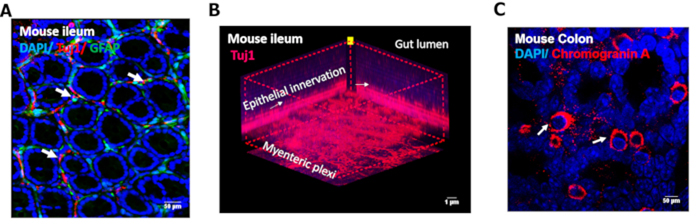
CLARITY (Clear Lipid-exchanged Acrylamide-hybridized Rigid Imaging compatible Tissue hYdrogel) has recently evolved as a valuable technique involving acrylamide embedding to delipidate tissue (without sectioning) and to preserve the 3-D tissue structure for immunostaining. The technique is highly relevant in imaging the dynamic gut environment where different cell types interact during homeostasis and disease states. This method optimized for the mouse gut is described here, which helps to trace cell types like epithelia, enteroendocrine, neurons, glia, and the neuronal projections into the epithelia or enteroendocrine cells that mediate microbial sensing or nutrient chemo sensing respectively. The gut tissue (1-1.5 cm) is fixed in 4% paraformaldehyde (PFA) in phosphate buffered saline (PBS) at 4 °C overnight on day 1. On day 2, PFA is discarded, and the tissue is washed thrice with PBS. The tissue is hydrogel embedded to preserve its integrity by incubation in 4% hydrogel (acrylamide) solution in PBS (diluted from 30% ProtoGel) overnight at 4 °C. On day 3, the tissue-hydrogel solution is incubated at 37 °C for 1 h to allow hydrogel polymerization. Tissue is then washed thrice gently with PBS to remove excess hydrogel. The subsequent step of delipidation (clearing) involves tissue incubation in sodium dodecyl sulfate (8% SDS in PBS) at 37 °C for 2 days (days 4 & 5) on a shaker at room temperature (RT). On day 6, the cleared tissue is thoroughly washed with PBS to remove SDS. Tissue can be immunostained by incubation in primary antibodies (diluted in 0.5% normal donkey serum in PBS containing 0.3% Triton X-100), overnight at 4°C, and subsequent incubation in appropriate secondary Alexa Fluor antibodies for 1.5 h at RT, and nuclear staining with DAPI (1: 10000). The tissue is transferred onto a clean glass slide and mounted using VectaShield for confocal imaging.
Figures
References
Publication types
MeSH terms
Substances
Grants and funding
LinkOut - more resources
Full Text Sources
