. 2021 Jul 21;49(13):7644-7664.
doi: 10.1093/nar/gkab508.

Assembly of higher-order SMN oligomers is essential for metazoan viability and requires an exposed structural motif present in the YG zipper dimer

Affiliations

Assembly of higher-order SMN oligomers is essential for metazoan viability and requires an exposed structural motif present in the YG zipper dimer

Kushol Gupta et al. Nucleic Acids Res. .

Abstract

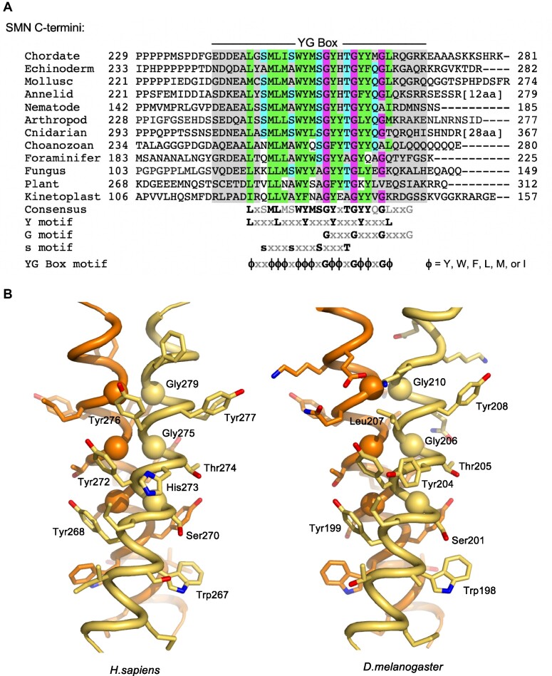
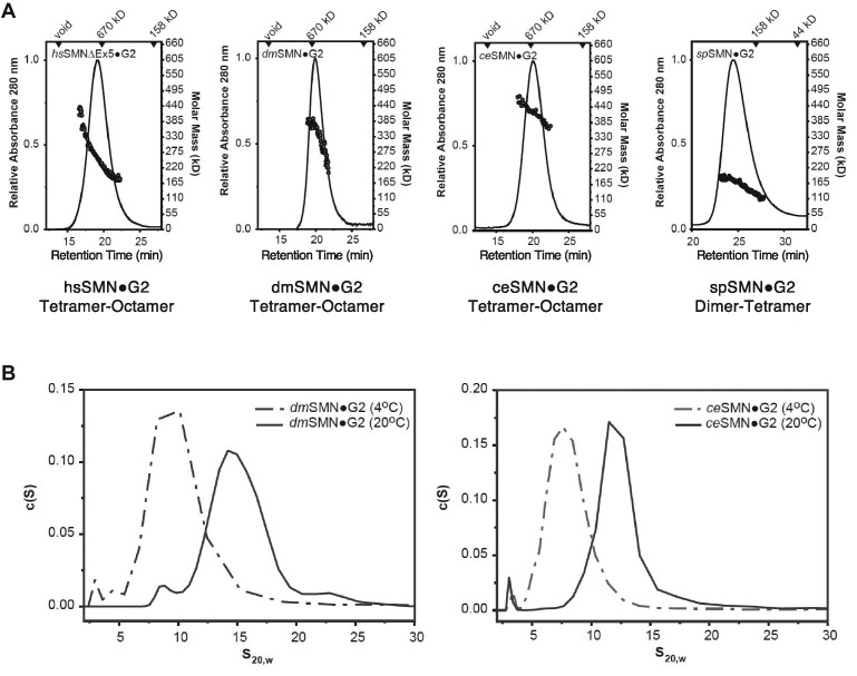
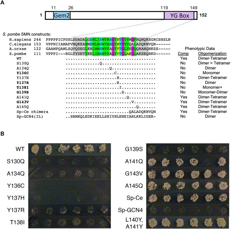
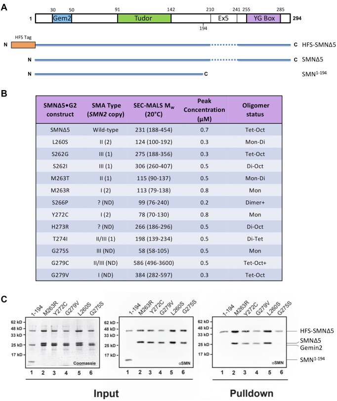
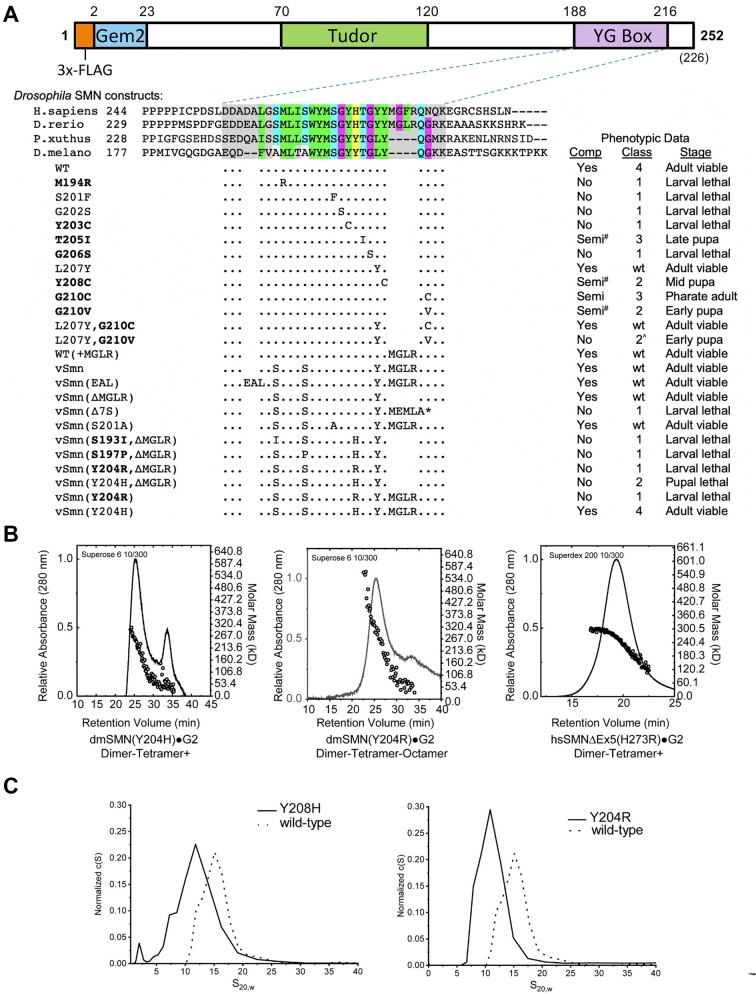
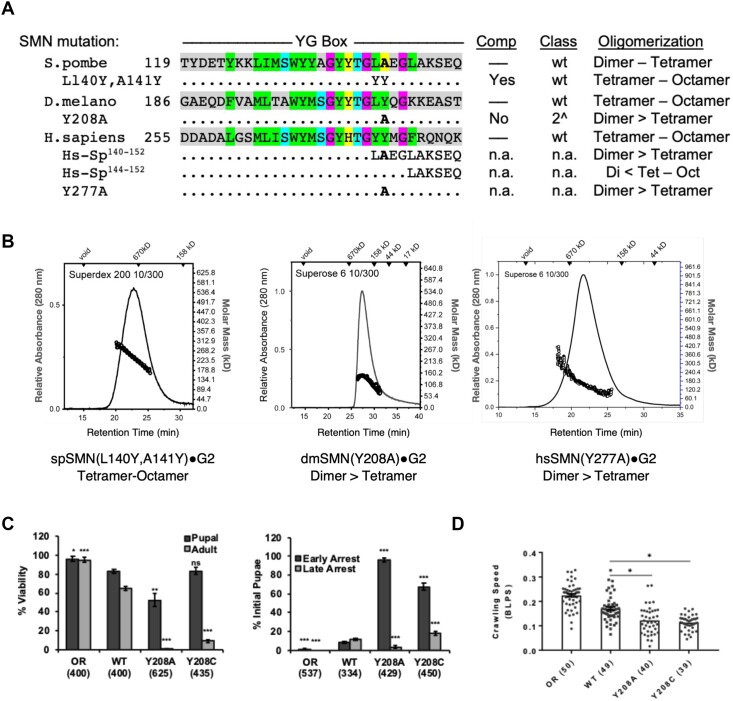
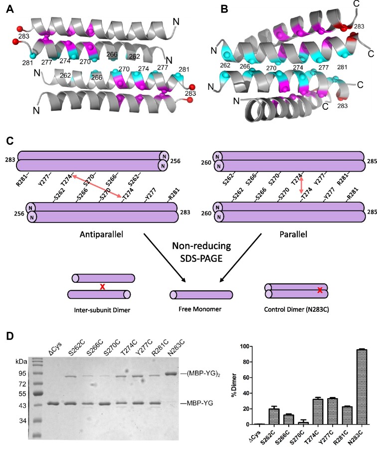
Protein oligomerization is one mechanism by which homogenous solutions can separate into distinct liquid phases, enabling assembly of membraneless organelles. Survival Motor Neuron (SMN) is the eponymous component of a large macromolecular complex that chaperones biogenesis of eukaryotic ribonucleoproteins and localizes to distinct membraneless organelles in both the nucleus and cytoplasm. SMN forms the oligomeric core of this complex, and missense mutations within its YG box domain are known to cause Spinal Muscular Atrophy (SMA). The SMN YG box utilizes a unique variant of the glycine zipper motif to form dimers, but the mechanism of higher-order oligomerization remains unknown. Here, we use a combination of molecular genetic, phylogenetic, biophysical, biochemical and computational approaches to show that formation of higher-order SMN oligomers depends on a set of YG box residues that are not involved in dimerization. Mutation of key residues within this new structural motif restricts assembly of SMN to dimers and causes locomotor dysfunction and viability defects in animal models.


Figures

Figure 1.
Figure 1.
Structural conservation of the SMN YG box domain. (A) Phylogenetic analysis of SMN C-termini from a diverse selection of eukaryotes. Conserved glycine residues are shaded in magenta, hydrophobic residues are in green, and polar residues in teal. Note the regularized spacing of residues in three overlapping motifs (Y, G and s) that are contained within the overall YG box consensus. (B) Structure of the SMN YG box dimer. Experimental atomic structures of the human (PDB ID: 4GLI) and fission yeast (PDB ID: 4RG5) SMN dimers were used to generate extended models of the human (left) and fruitfly (right) proteins. The three well-conserved glycine residues are shown as Cα spheres. Atomic views were rendered using the program PYMOL (73). See supplemental Supplementary Figure S1 for a more extensive phylogenetic comparison along with additional ultrastructural models.
Figure 2.
Figure 2.
Biophysical properties of wild-type SMN•Gemin2 complexes are conserved. (A) SEC-MALS analysis performed at 20°C for Homo sapiens (hs), Drosophila melanogaster (dm), Caenorhabditis elegans (ce) and Schizosaccharomyces pombe (sp) SMN•G2 complexes. As previously observed for the human and fission yeast complexes (24,25), SEC elution times suggest the presence of larger multimers in metazoans. However, Mw values from the in-line light scattering indicate a range of oligomers in each case; the oligomeric range is assigned below each panel. (B) Sedimentation velocity (SV-AUC) analyses of dmSMN•G2 (left) and ceSMN•G2 (right). At 25°C, the fruitfly complex sediments as a broad ∼15 S peak but at 4°C a smaller ∼8 S peak is observed. Similarly, the nematode complex sediments as a broad ∼12S peak at 25°C but smaller ∼7S peak at 4°C. See Supplementary Figure S2 for additional biophysical characterization of the dmSMN complex and its components.
Figure 3.
Figure 3.
Genetic and biophysical characterization of fission yeast SMN mutants. (A) Cartoon of spSMN protein, showing relative location of the Gemin2 (Gem2) binding domain and the YG box. An alignment of YG box sequences from the human (H. sapiens), nematode (C. elegans), köji mold (A.orzae) and fission yeast (S. pombe) SMN orthologs is shown for comparison. Genetic complementation analysis of a fission yeast smn1 null allele was performed with a wild-type (WT) rescue construct or with a variety of chimeric or point substitution mutation constructs. Mutants that correspond to human SMA-causing missense alleles are shown in bold text. Ability to complement (Comp) the growth defect observed in the null mutant background is indicated. Recombinant SMN•G2 complexes of these same mutant constructs were generated in vitro and subjected to SEC-MALS and SE-AUC analysis, as described in Figure 2. The range of oligomeric species detected by SEC-MALS is also indicated. See Supplementary Table S2 for additional details regarding biophysical characterization of spSMN complexes. (B) Complementation of smn1+ deletion in S. pombe. Constructs expressing smn1 variants under control of the nmt1 promoter were transformed into haploid S. pombe containing a deletion of chromosomal smn1+ and episomal smn1 expressed from a ura4+ plasmid. Individual transformants were patched onto minimal medium containing thiamine, then replica plated to dilution on selection plates containing 5-fluoroorotic acid (5-FOA). Strong growth on FOA medium requires loss of the ura4+ plasmid, and therefore complementation of smn1Δ by the smn construct.
Figure 4.
Figure 4.
Biophysical characterization of human SMN•Gemin2 complexes bearing SMA-causing YG box missense mutations. (A) Cartoon of hsSMN protein, showing the conserved YG box, Gemin2 binding (Gem2) and Tudor domains, along with location of exon5 (Ex5) sequences deleted in the SMNΔ5 construct. (B) SEC-MALS analysis of SMA-causing point mutant constructs in the hsSMNΔ5•Gemin2 backbone. All of the mutations were generated on the SMNΔ5 backbone. Additional details regarding reported human SMN2 copy number and SMA patient phenotype are also provided (22). (C) Formation of mixed oligomers between wild-type hsSMN and SMA patient mutations in vitro. A subset of the patient-derived mutations was screened for the ability to form mixed oligomers with wild-type SMNΔ5. The left two panels show Coomassie and western blot analyses of SDS-PAGE gels of the input material following bacterial co-expression and lysate clarification; the last panel shows a western blot of the resulting pulldown using Chitin-binding resin. As a negative control, the ability of a truncated SMN lacking the YG oligomerization domain (SMN1–194) was also assayed. After co-expression and elution from the chitin binding resin, four of the five patient mutant samples (M263R, Y272C, G279V and L260S) demonstrated the ability to form mixed oligomers. Among these five missense constructs, only SMNΔ5(G275S) failed to co-purify with wild-type SMNΔ5.
Figure 5.
Figure 5.
Genetic and biophysical characterization of Smn YG box mutations in Drosophila. (A) Cartoon of the dmSMN protein, showing the conserved YG box, Gemin2 binding (Gem2) and Tudor domains, along with the location of the 3x-FLAG tag used for transgenic rescue experiments. An alignment of YG box sequences from the human (H.sapiens), zebrafish (D.rerio), butterfly (P.xuthus) and fruitfly (D.melanogaster) SMN orthologs is shown for reference. Phenotypic comparisons of an extensive panel of Drosophila YG box substitution mutations are summarized below the alignment. With the exception of S201F and G202S, which are point mutations in the endogenous Smn gene (55), the rest of the panel is comprised of transgenic constructs that have been recombined with an Smn null allele ((21); this work). Genetic complementation analysis (Comp) was performed using either a wild-type (WT) or mutant Flag-Smn transgene. Mutants that correspond to human SMA-causing missense alleles are shown in bold text; the classification system used for fly SMA models was described previously (44). See Supplementary Figure S4 for pupal and adult viability analysis of the fifteen new transgenic fly lines used in this work. Mutant lines that eclose at low frequency (adult escapers) are considered semi-viable; those marked with a # sign are incapable of establishing an independent breeding stock. Class 2^ denotes animals that pupate but display a significant advancement in lethal phase or reduction in eclosion frequency. The asterisk * = stop codon. (B) SEC-MALS analysis of SMN•G2 complexes containing mutations at hsH273 (H273R) or dmY204 (Y204H and Y204R) aimed at modeling an SMA-causing missense allele in human SMN1. (C) Analytical ultracentrifugation of Drosophila SMN•G2 complexes. c(S) sedimentation distributions of wild-type (WT) or mutant (Y204H and Y204R) are shown. Note, biophysical analyses of additional Drosophila SMN constructs are presented in Table 1 and Supplementary Figure S2.
Figure 6.
Figure 6.
Identification of a specific YG box residue critical for formation of higher-order SMN multimers. A structure-function analysis was carried out in parallel using fission yeast, fruitfly and human SMN. (A) Genetic complementation (Comp) and biophysical analyses (Oligomerization) of C-terminal chimeras and point substitution mutants. (B) SEC-MALS analysis of SMN•G2 complexes of yeast spSMNL140Y,A141Y, dmSMNY208A and hsSMNY277A. (C) Developmental viability of control Oregon Red (OR) flies or animals expressing transgenic Flag-tagged Smn wild-type (WT), Y208C or Y208A rescue constructs in the background of an Smn null mutation. Left panel: % Viability is the proportion of animals that survive to the pupal (darker gray) or adult (lighter gray) stages, relative to the number of larvae initially collected (n-values in parentheses). Right panel: Breakdown of fraction of animals that arrest during early versus late stages of pupal development. (D) Larval locomotion analysis. Average crawling speed, measured in body lengths/sec, in wandering third instar larvae. Data points correspond to measurements of individual larvae. n-values for each genotype are shown in parentheses. Statistical analysis used in panels A–D: Asterisks above the data bars indicate significance versus WT rescue line from one-way ANOVA using the Dunnet correction for multiple comparisons. *P < 0.05, **P < 0.01, ***P < 0.001. P > 0.05 is not significant (ns).
Figure 7.
Figure 7.
Disulfide crosslinking analysis and models of higher order SMN oligomerization. (A) Model of an antiparallel SMN YG box tetramer based on crystal contacts in a spSMN crystal structure (PDB ID: 7BB3). Numbering as per human SMN. (B) Model of a parallel YG box tetramer, based on s-motif helix–helix interactions observed in membrane proteins (see Materials and Methods and text for details). (C) Cartoon of disulfide crosslinking between YG box dimers containing single cysteine substitutions at indicated residues. Crosslinking between Thr274 residues is illustrated with a red arrow as an example. Disulfide crosslinking allows for resolution of dimers upon non-reducing SDS-PAGE. The N283C substitution serves as a positive control because hsN283 (marked in red in panels A and B) is within the dimer interface and is efficiently crosslinked. (D) SDS-PAGE of MBP-hsSMN YG box fusions treated with 160 μM diamide for 60 min (left). The graph on the right shows the average fraction of dimers formed from three independent crosslinking experiments. Error bars show standard deviation. Note that MBP-YG box fusions treated with 1 mM DTT rather than diamide are monomeric (see Supplementary Figure S8).

References

    1. Levy E.D., Pereira-Leal J.B., Chothia C., Teichmann S.A.. 3D complex: a structural classification of protein complexes. PLoS Comput. Biol. 2006; 2:e155. - PMC - PubMed
    1. Liu H., Cao M., Wang Y., Lv B., Li C.. Bioengineering oligomerization and monomerization of enzymes: learning from natural evolution to matching the demands for industrial applications. Crit. Rev. Biotechnol. 2020; 40:231–246. - PubMed
    1. Ali M.H., Imperiali B.. Protein oligomerization: how and why. Bioorg. Med. Chem. 2005; 13:5013–5020. - PubMed
    1. Matera A.G., Wang Z.. A day in the life of the spliceosome. Nat. Rev. Mol. Cell Biol. 2014; 15:108–121. - PMC - PubMed
    1. Gruss O.J., Meduri R., Schilling M., Fischer U.. UsnRNP biogenesis: mechanisms and regulation. Chromosoma. 2017; 126:577–593. - PubMed

Publication types

Substances

LinkOut - more resources