. 2021 Aug 17;120(16):3437-3454.
doi: 10.1016/j.bpj.2021.05.027. Epub 2021 Jun 25.

Factors underlying asymmetric pore dynamics of disaggregase and microtubule-severing AAA+ machines

Affiliations

Factors underlying asymmetric pore dynamics of disaggregase and microtubule-severing AAA+ machines

Mangesh Damre et al. Biophys J. .

Abstract

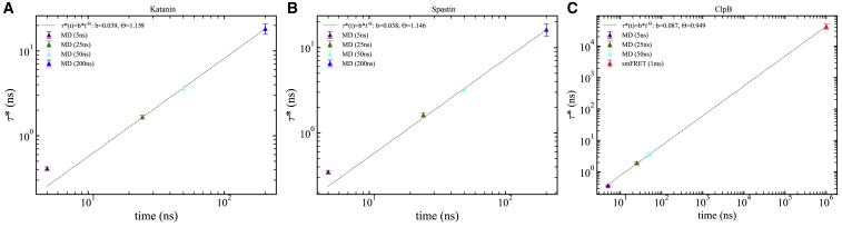
Disaggregation and microtubule-severing nanomachines from the AAA+ (ATPases associated with various cellular activities) superfamily assemble into ring-shaped hexamers that enable protein remodeling by coupling large-scale conformational changes with application of mechanical forces within a central pore by loops protruding within the pore. We probed the asymmetric pore motions and intraring interactions that support them by performing extensive molecular dynamics simulations of single-ring severing proteins and the double-ring disaggregase ClpB. Simulations reveal that dynamic stability of hexameric pores of severing proteins and of the nucleotide-binding domain 1 (NBD1) ring of ClpB, which belong to the same clade, involves a network of salt bridges that connect conserved motifs of central pore loops. Clustering analysis of ClpB highlights correlated motions of domains of neighboring protomers supporting strong interprotomer collaboration. Severing proteins have weaker interprotomer coupling and stronger intraprotomer stabilization through salt bridges involving pore loops. Distinct mechanisms are identified in the NBD2 ring of ClpB involving weaker interprotomer coupling through salt bridges formed by noncanonical loops and stronger intraprotomer coupling. Analysis of collective motions of PL1 loops indicates that the largest amplitude motions in the spiral complex of spastin and ClpB involve axial excursions of the loops, whereas for katanin they involve opening and closing of the central pore. All three motors execute primarily axial excursions in the ring complex. These results suggest distinct substrate processing mechanisms of remodeling and translocation by ClpB and spastin compared to katanin, thus providing dynamic support for the differential action of the two severing proteins. Relaxation dynamics of the distance between the PL1 loops and the center of mass of protomers reveals observation-time-dependent dynamics, leading to predicted relaxation times of tens to hundreds of microseconds on millisecond experimental timescales. For ClpB, the predicted relaxation time is in excellent agreement with the extracted time from smFRET experiments.


Figures

Figure 1
Figure 1
Structural details of the hexamer of severing proteins and ClpB disaggregase. Katanin spiral conformation (PDB: 6UGD): (a) hexamer (top view); (b) PL1 loops. Relaxation time analysis focuses on the distance between each R267 residue (small beads) of PL1 loops and the COM of the corresponding protomer (large beads, blue); (c) transparent surface highlighting the HBD (red) and NBD (blue) domains in a protomer; (d) side view of spiral arrangement of PL1 (blue), PL2 (red), and PL3 (green) loops and the bound substrate (E14) in the central pore (in orange surface). ClpB spiral conformation (PDB: 6OAY): (e) hexamer (top view); (f) PL1 loops. Distance between each R252 residue (small beads) of PL1 loops and the COM of each protomer (large beads, blue); (g) transparent surface highlighting the NBD1 (blue) and NBD2 (red) domains in a protomer; (h) side view of spiral arrangement of PL1 (blue) and PL3 (green) loops and the bound substrate (polyA) in the central pore (in orange surface). All molecular structures shown in this work were created using Visual Molecular Dynamics (51). To see this figure in color, go online.
Figure 2
Figure 2
Principal collective motions of pore loops of severing proteins and ClpB. Motions corresponding to principal components PC1 and PC2 are shown for the spiral state of (ad) katanin (6UGD) in (a and b) the nucleotide- and substrate-free (APO) configuration and (c and d) the nucleotide- and substrate-bound (ATP + E14) configuration; (eh) spastin (6PO7) in (e and f) the nucleotide- and substrate-free (APO) configuration and (g and h) the nucleotide- and substrate-bound (ATP + E15) configuration; and (il) ClpB (6OAY) in (i and j) the nucleotide- and substrate-free (APO) configuration and (k and l) the nucleotide- and substrate-bound (ATP + ALA) configuration. Pore loops of individual protomers are color coded, following the convention from Fig. 1, and arrows indicate directions and relative amplitudes of amino acid motions. The pore axis is oriented parallel to the z axis. Fig. S5 displays corresponding motions for ring states. To see this figure in color, go online.
Figure 3
Figure 3
Interprotomer cluster patterns of severing proteins and ClpB. (a) Katanin spiral ATP state: coupling between HBD domains of seam protomers, A and F, and the NBD domain of protomer B. (b) ClpB spiral ALA + ATP state: coupling between large (L) and small (S) domains of NBD1 domains of neighboring protomers in the counterclockwise direction. Protomers are color coded, and clusters are highlighted using a transparent surface (red). To see this figure in color, go online.
Figure 4
Figure 4
Networks of complex salt bridges identified in severing proteins and ClpB disaggregase. Salt bridge networks identified in our simulations in (a) katanin spiral (6UGD) and ring (6UGE) states, highlighted in pink and cyan; (b) spastin spiral (6P07) in cyan; (c) spastin ring (6PEN) in cyan; and (d) NBD1 of ClpB in spiral (6OAY) and ring (6OAX) states in pink are shown. Networks include complex salt bridges (dashes), in which specific charged residues (indicated using stick representation) may participate in multiple interaction pairs and couple at least three protomer pairs. Highlighted regions illustrate salt bridges that form at one interprotomer interface. Protomers are color coded, and the substrate peptide is shown in gray or green. The detailed list of salt bridges formed is indicated in the text and in Tables S5 and S6. To see this figure in color, go online.
Figure 5
Figure 5
Dependence of relaxation time (τ) on the observation timescale. Log-log plots indicate power-law dependence (dashed line) of τ versus time (nanoseconds) for (A) katanin (MD data) and (B) spastin (MD data) and (C) ClpB (MD + smFRET data). Data from MD simulations of 5 ns (magenta), 25 ns (green), 50 ns (cyan), and 200 ns (blue) trajectories, as well as from smFRET experiments (red) (70) are shown. Standard error of τ data is shown as error bars. To see this figure in color, go online.

References

    1. Glover J.R., Lindquist S. Hsp104, Hsp70, and Hsp40: a novel chaperone system that rescues previously aggregated proteins. Cell. 1998;94:73–82. - PubMed
    1. Erzberger J.P., Berger J.M. Evolutionary relationships and structural mechanisms of AAA+ proteins. Annu. Rev. Biophys. Biomol. Struct. 2006;35:93–114. - PubMed
    1. Doyle S.M., Wickner S. Hsp104 and ClpB: protein disaggregating machines. Trends Biochem. Sci. 2009;34:40–48. - PubMed
    1. Martin A., Baker T.A., Sauer R.T. Pore loops of the AAA+ ClpX machine grip substrates to drive translocation and unfolding. Nat. Struct. Mol. Biol. 2008;15:1147–1151. - PMC - PubMed
    1. Lee S., Sowa M.E., Tsai F.T. The ClpB/Hsp104 molecular chaperone-a protein disaggregating machine. J. Struct. Biol. 2004;146:99–105. - PubMed

Publication types

LinkOut - more resources