Review
. 2021 Oct:478:102-121.
doi: 10.1016/j.ydbio.2021.06.012. Epub 2021 Jun 25.

Human neural organoids: Models for developmental neurobiology and disease

Affiliations
Review

Human neural organoids: Models for developmental neurobiology and disease

Brian Guy et al. Dev Biol. 2021 Oct.

Abstract

Human organoids stand at the forefront of basic and translational research, providing experimentally tractable systems to study human development and disease. These stem cell-derived, in vitro cultures can generate a multitude of tissue and organ types, including distinct brain regions and sensory systems. Neural organoid systems have provided fundamental insights into molecular mechanisms governing cell fate specification and neural circuit assembly and serve as promising tools for drug discovery and understanding disease pathogenesis. In this review, we discuss several human neural organoid systems, how they are generated, advances in 3D imaging and bioengineering, and the impact of organoid studies on our understanding of the human nervous system.

Keywords: Brain; CRISPR; Cerebellar organoid; Cerebellum; Cerebral cortex; Cortical organoid; Embryoid bodies; Embryonic stem cells; Hippocampal organoid; Hippocampus; Human; Hypothalamic organoid; Hypothalamus; Induced-pluripotent stem cells; Organoid; Retina; Retinal organoid; Thalamic organoid; Thalamus; scRNA-seq.


Figures

Figure 1.
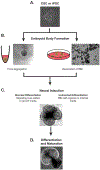
Figure 1.. Organoid differentiation strategies.
Representative images of different stages of retinal organoid differentiation. A) Embryonic or induced pluripotent stem cells are cultured in 2D. B) Two methods of embryoid body formation have been developed. Left: aggregation by gravity of stem cells. Right: dissociation of embryoid bodies from 2D culture. C) Neural induction: embryoid bodies are driven to neuroectodermal fate by directed or undirected differentiation. D) Differentiation and Maturation: Following neural induction, organoids are differentiated into specific neural tissue types, exhibiting developmental, morphological, and cytological similarities to in vivo tissues (Benito-Kwiecinski and Lancaster, 2020; Eiraku et al., 2011; Eiraku et al., 2008; Eldred et al., 2018; Lancaster and Knoblich, 2014; Schutgens and Clevers, 2020; Wang, 2018; Xu et al., 2018).
Figure 2.
Figure 2.. Schematic of brain vesicle differentiation during neural tube development.
A) The neural tube divides into three primary brain vesicles: prosencephalon (forebrain), mesencephalon (midbrain), and rhombencephalon (hindbrain). At the secondary vesicle stage, the prosencephalon subdivides into the telencephalon and diencephalon, whereas the rhombencephalon subdivides into the metencephalon and myelencephalon. B) Diagram of the embryonic brain regions and mature brain structures. Abbreviate: Telencephalon (Telen), Diencephalon (Dien), Mesencephalon (Mesen), Metencephalon (Meten), Myelencephalon (Myelen), and Spinal Cord (S.C)(Allen, 2008; Kiecker and Lumsden, 2005; Martinez-Ferre and Martinez, 2012; Shimogori et al., 2004; Stiles and Jernigan, 2010).
Figure 3.
Figure 3.. Regional localization of extrinsic and intrinsic factors in the vertebrate neural tube during development.
A) Localization of extrinsic factors and B) intrinsic factors. Abbreviate: Bone Morphogenic Proteins (BMPs), Fibroblast Growth Factors (FGFs), Retinoic Acid (RA), Sonic Hedgehog (SHH), and Wingless (WNTs). (Allen, 2008; Allodi and Hedlund, 2014; Battiste et al., 2007; Boncinelli et al., 1993; Chen et al., 2005; Dono et al., 2002; Echelard et al., 1994; Ellis et al., 2004; Furukawa et al., 1997; Jones et al., 2002; Kataoka and Shimogori, 2008; Kim et al., 2021; Lee et al., 2011; Lewis et al., 2008; Martin et al., 2012; Martinez Barbera et al., 2000; Martinez-Ferre and Martinez, 2009, 2012; Maruoka et al., 1998; Oliver et al., 1995; Perez-Balaguer et al., 2009; Shimogori et al., 2004; Waters et al., 2003; Winnier et al., 1995).
Figure 4.
Figure 4.. Organoid differentiation protocols.
Table indicates brain organoid, representative image of each organoid, method for neural induction, method for differentiation and maturation, and source. Cerebral cortex organoid image from Lancaster et al (2013). Hippocampal organoid image from Sakaguchi et al (2015). Retinal organoid image from Kruczek et al (2021). Thalamic organoid image from Xiang et al (2019). Hypothalamic and midbrain organoid images from Qian et al (2016). Cerebellum organoid image from Muguruma et al 2015(Bhaduri et al., 2020; Jo et al., 2016; Kadoshima et al., 2013; Kruczek and Swaroop, 2020; Lancaster et al., 2013; Meyer et al., 2009; Muguruma et al., 2015; Pollen et al., 2019; Qian et al., 2016; Sakaguchi et al., 2015; Sloan et al., 2017; Trujillo et al., 2019; Wahlin et al., 2017; Xiang et al., 2017; Zhong et al., 2014).

References

    1. Adijanto J, Castorino JJ, Wang ZX, Maminishkis A, Grunwald GB, and Philp NJ (2012). Microphthalmia-associated transcription factor (MITF) promotes differentiation of human retinal pigment epithelium (RPE) by regulating microRNAs-204/211 expression. J Biol Chem 287, 20491–20503. - PMC - PubMed
    1. Agirman G, Broix L, and Nguyen L (2017). Cerebral cortex development: an outside-in perspective. FEBS Lett 591, 3978–3992. - PubMed
    1. Alladin A, Chaible L, Garcia Del Valle L, Sabine R, Loeschinger M, Wachsmuth M, Heriche JK, Tischer C, and Jechlinger M (2020). Tracking cells in epithelial acini by light sheet microscopy reveals proximity effects in breast cancer initiation. Elife 9. - PMC - PubMed
    1. Allen I (2008). Allen Developing Mouse Brain Atlas.
    1. Allodi I, and Hedlund E (2014). Directed midbrain and spinal cord neurogenesis from pluripotent stem cells to model development and disease in a dish. Front Neurosci 8, 109. - PMC - PubMed

Publication types