. 2021:31:102728.
doi: 10.1016/j.nicl.2021.102728. Epub 2021 Jun 17.

Typical somatomotor physiology of the hand is preserved in a patient with an amputated arm: An ECoG case study

Affiliations

Typical somatomotor physiology of the hand is preserved in a patient with an amputated arm: An ECoG case study

Max van den Boom et al. Neuroimage Clin. 2021.

Abstract

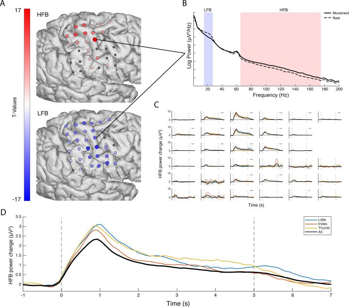
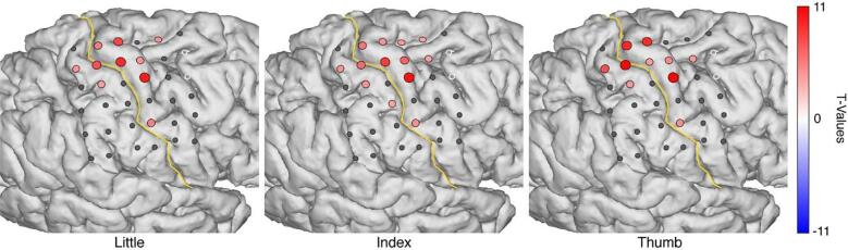
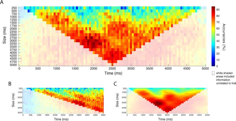
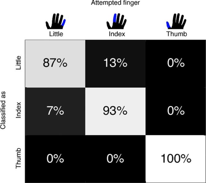
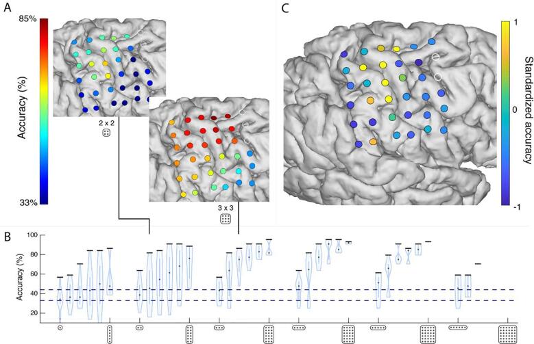
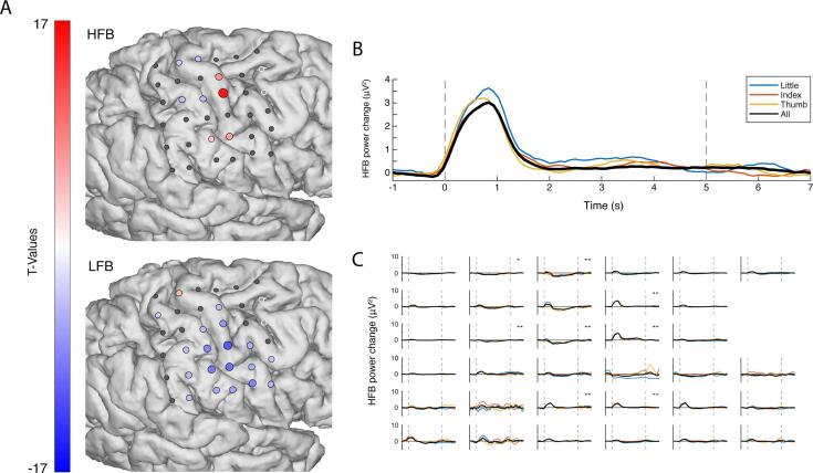
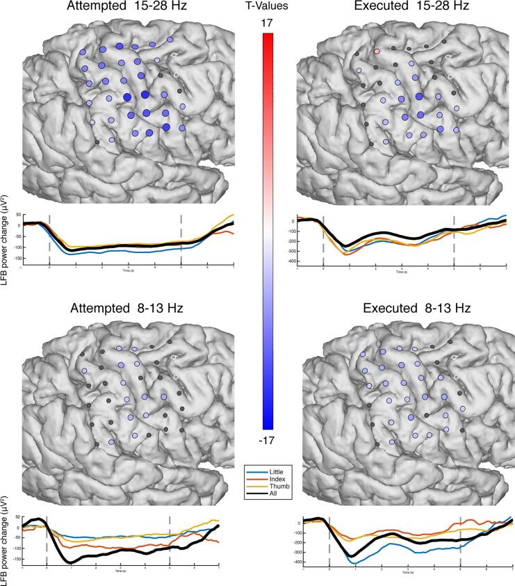
Electrophysiological signals in the human motor system may change in different ways after deafferentation, with some studies emphasizing reorganization while others propose retained physiology. Understanding whether motor electrophysiology is retained over longer periods of time can be invaluable for patients with paralysis (e.g. ALS or brainstem stroke) when signals from sensorimotor areas may be used for communication or control over neural prosthetic devices. In addition, a maintained electrophysiology can potentially benefit the treatment of phantom limb pains through prolonged use of these signals in a brain-machine interface (BCI). Here, we were presented with the unique opportunity to investigate the physiology of the sensorimotor cortex in a patient with an amputated arm using electrocorticographic (ECoG) measurements. While implanted with an ECoG grid for clinical evaluation of electrical stimulation for phantom limb pain, the patient performed attempted finger movements with the contralateral (lost) hand and executed finger movements with the ipsilateral (healthy) hand. The electrophysiology of the sensorimotor cortex contralateral to the amputated hand remained very similar to that of hand movement in healthy people, with a spatially focused increase of high-frequency band (65-175 Hz; HFB) power over the hand region and a distributed decrease in low-frequency band (15-28 Hz; LFB) power. The representation of the three different fingers (thumb, index and little) remained intact and HFB patterns could be decoded using support vector learning at single-trial classification accuracies of >90%, based on the first 1-3 s of the HFB response. These results indicate that hand representations are largely retained in the motor cortex. The intact physiological response of the amputated hand, the high distinguishability of the fingers and fast temporal peak are encouraging for neural prosthetic devices that target the sensorimotor cortex.

Keywords: BCI; ECoG; Limb loss; Motor cortex; Physiology; Upper arm amputation.


Figures

Fig. 1
Fig. 1
The electrophysiological response during attempted movement versus rest. (A). The changes in HFB (top, 65–175 Hz) and LFB (bottom, 15–28 Hz) for each grid electrode. Electrodes with a red or blue color had a significant change in band-power, whereas electrodes with an insignificant change in band-power are shown in grey; the two excluded electrodes are shown in white. (B) The power spectra of movement (solid line) and rest (dashed line) for a single electrode. (C) The HFB power changes over time, each graph represents one electrode. The black line represents all fingers, whereas the colored lines represent individual fingers. The two vertical dotted lines indicated the cue on- and offset. (D) The HFB power changes over time were averaged across those electrodes that showed a significant increase for a condition (blue: little, red: index, yellow: thumb, black: all fingers). The black trace has a lower amplitude because a different set of significant electrodes contributed to each trace, with more significant, but lower amplitude electrodes contributing to the black trace. (For interpretation of the references to color in this figure legend, the reader is referred to the web version of this article.)
Fig. 2
Fig. 2
The HFB power changes upon attempted movement of each individual finger. Electrodes with a significant change in HFB band-power are shown in red, whereas electrodes with an insignificant HFB change are shown in grey, the two electrodes that were excluded are shown in white. The yellow line represents the central sulcus. (For interpretation of the references to color in this figure legend, the reader is referred to the web version of this article.)
Fig. 3
Fig. 3
The classification accuracies at different window sizes (y-axes) and offsets from cue onset (x-axes), based on half of the data (~45 trials). (A) The x-axis indicates the center of each window. (B) The same plot shown, but with the x-axis indicating the right of each window, such that each time point includes the information present before that time. (C) Shows the classification accuracies smoothed with a Gaussian filter (offset σ: 0.5, size σ: 2.5). The white-shaded regions in each graph indicate the classification accuracies in which the window included information unrelated to the trial (i.e. rest before or after the trial).
Fig. 4
Fig. 4
Confusion matrix with the classification scores of the individual fingers. Each column represents the finger of which movement was attempted. The rows represent how each of those finger movements was classified.
Fig. 5
Fig. 5
The spatial distribution of information. (A) Searchlight classification maps of searchlight with 2 × 2 (top) and 3 × 3 electrodes (bottom). (B) The classification results of the different searchlights, ranging from 1 × 6 to 6 × 6 grids in two directions (superior-inferior, anterior-posterior). Each violin plot represents the searchlight results with a specific grid configuration. The violin represents the distribution of the classification accuracies at the different searchlight positions within the grid, with a black horizontal bar to indicate the searchlight position that classified the highest. The lower dotted blue line shows the chance level at 33%, while the upper blue line indicates the threshold of 45% above which the decoding accuracy was significant. Note that, for the larger searchlight grids, virtual placements were limited or not possible due to overlap with the bad electrodes. (C) Most informative electrodes, identified by a random search classification on 10.000 subsets of electrodes. (For interpretation of the references to color in this figure legend, the reader is referred to the web version of this article.)
Fig. 6
Fig. 6
The, ipsilateral, electrophysiological response of executed movement versus rest. (A) The changes in HFB (top, 65–175 Hz) and LFB power (bottom, 15–28 Hz) for each grid electrode. Electrodes with a red or blue color showed a significant change in band-power, whereas electrodes with an insignificant change in band-power are shown in grey; the two excluded electrodes are shown in white. (B) HFB power changes over time averaged across those electrodes that showed a significant positive increase. The black line represents all fingers, whereas the colored lines represent individual fingers. The two vertical dotted lines indicated the cue on- and offset. (C) The HFB power changes over time, each graph represents one electrode. (For interpretation of the references to color in this figure legend, the reader is referred to the web version of this article.)
Supplementary figure 1

Similar articles

Cited by

References

    1. Bai O., Lin P., Huang D., Fei D.Y., Floeter M.K. Towards a user-friendly brain-computer interface: initial tests in ALS and PLS patients. Clin. Neurophysiol. 2010;121:1293–1303. - PMC - PubMed
    1. Benabid A.L., Costecalde T., Eliseyev A., Charvet G., Verney A., Karakas S., Foerster M., Lambert A., Morinière B., Abroug N., Schaeffer M.C., Moly A., Sauter-Starace F., Ratel D., Moro C., Torres-Martinez N., Langar L., Oddoux M., Polosan M., Pezzani S., Auboiroux V., Aksenova T., Mestais C., Chabardes S. An exoskeleton controlled by an epidural wireless brain–machine interface in a tetraplegic patient: a proof-of-concept demonstration. Lancet Neurol. 2019;18:1112–1122. - PubMed
    1. Birbaumer N., Lutzenberger W., Montoya P., Larbig W., Unertl K., Töpfner S., Grodd W., Taub E., Flor H. Effects of regional anesthesia on phantom limb pain are mirrored in changes in cortical reorganization. J. Neurosci. 1997;17:5503–5508. - PMC - PubMed
    1. Bishop C.M. Springer-Verlag; New York: 2006. Machine Learning and Pattern Recoginiton, Information Science and Statistics.
    1. Blakely T.M., Olson J.D., Miller K.J., Rao R.P.N., Ojemann J.G. Neural correlates of learning in an electrocorticographic motor-imagery brain-computer interface. Brain-Comput. Interfaces. 2014;1:147–157. - PMC - PubMed

Publication types