

Integrin α5β1 Mediated Cellular Reorganization in Human Mesenchymal Stem Cells During Neuronal Differentiation
- PMID: 34182488
- PMCID: PMC8286471
- DOI: 10.21873/invivo.12482
Integrin α5β1 Mediated Cellular Reorganization in Human Mesenchymal Stem Cells During Neuronal Differentiation
Abstract
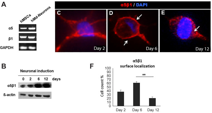
Background/aim: Mesenchymal stem cells (MSCs) have been widely used for yielding neurons in culture to study nervous system pathologies and develop regenerative approaches. In this study, cellular rearrangements of human MSCs related to the expression of the fibronectin common receptor integrin α5β1 and its cell surface localization during neuronal differentiation, were examined.
Materials and methods: Proliferation kinetics of neuronal induced hMSCs (hMd-Neurons) were quantified by BrdU assay, and hMd-Neurons were immunostained for neuronal marker expression. Additionally, cDNA and protein samples were collected at different time points for integrin α5β1 expression analysis.
Results: Endogenous integrin α5β1 expression was significantly upregulated by day 6 and maintained until day 12. Cell surface localization of α5β1 integrin was increased by day 6; the integrin was internalized into the cytosol by day 12.
Conclusion: Integrin dynamics around day 6 of differentiation might be involved in neuronal differentiation and maturation or specification of hMd-Neurons.
Keywords: Mesenchymal stem cells; integrin; integrin α5β1; neuronal differentiation.
Copyright © 2021 International Institute of Anticancer Research (Dr. George J. Delinasios), All rights reserved.
Conflict of interest statement
The Authors declare no conflicts of interest in relation to this study.
Figures


Similar articles
-
Fibronectin-mediated upregulation of α5β1 integrin and cell adhesion during differentiation of mouse embryonic stem cells.Cell Adh Migr. 2011 Jan-Feb;5(1):73-82. doi: 10.4161/cam.5.1.13704. Epub 2011 Jan 1. Cell Adh Migr. 2011. PMID: 20962574 Free PMC article.
-
Intrinsic Surface Effects of Tantalum and Titanium on Integrin α5β1/ ERK1/2 Pathway-Mediated Osteogenic Differentiation in Rat Bone Mesenchymal Stromal Cells.Cell Physiol Biochem. 2018;51(2):589-609. doi: 10.1159/000495280. Epub 2018 Nov 20. Cell Physiol Biochem. 2018. PMID: 30458456
-
Wnt/β-catenin signaling mediates osteoblast differentiation triggered by peptide-induced α5β1 integrin priming in mesenchymal skeletal cells.J Biol Chem. 2015 Mar 13;290(11):6903-12. doi: 10.1074/jbc.M114.621219. Epub 2015 Jan 28. J Biol Chem. 2015. PMID: 25631051 Free PMC article.
-
IL-1β mediated nanoscale surface clustering of integrin α5β1 regulates the adhesion of mesenchymal stem cells.Sci Rep. 2021 Mar 25;11(1):6890. doi: 10.1038/s41598-021-86315-x. Sci Rep. 2021. PMID: 33767269 Free PMC article.
-
[Effects of integrin on differentiation of mesenchymal stem cells].Sheng Li Xue Bao. 2017 Aug 25;69(4):498-508. Sheng Li Xue Bao. 2017. PMID: 28825109 Review. Chinese.
Cited by
-
ITGA5 induces mesenchymal transformation to promote gliomas progression via PI3K/AKT/mTORC1 signaling pathway.Sci Rep. 2025 Apr 19;15(1):13539. doi: 10.1038/s41598-025-98170-1. Sci Rep. 2025. PMID: 40253517 Free PMC article.
References
-
- Kadereit S, Deeds LS, Haynesworth SE, Koc ON, Kozik MM, Szekely E, Daum-Woods K, Goetchius GW, Fu P, Welniak LA, Murphy WJ, Laughlin MJ. Expansion of LTC-ICs and maintenance of p21 and BCL-2 expression in cord blood CD34(+)/CD38(-) early progenitors cultured over human MSCs as a feeder layer. Stem Cells. 2002;20(6):573–582. doi: 10.1634/stemcells.20-6-573. - DOI - PubMed
-
- Nancarrow-Lei R, Mafi P, Mafi R, Khan W. A systemic review of adult mesenchymal stem cell sources and their multilineage differentiation potential relevant to musculoskeletal tissue repair and regeneration. Curr Stem Cell Res Ther. 2017;12(8):601–610. doi: 10.2174/1574888X12666170608124303. - DOI - PubMed
MeSH terms
Substances
LinkOut - more resources
Full Text Sources
Research Materials