. 2021 Jun 28;13(12):16816-16833.
doi: 10.18632/aging.203221. Epub 2021 Jun 28.

Celastrol protects against early brain injury after subarachnoid hemorrhage in rats through alleviating blood-brain barrier disruption and blocking necroptosis

Affiliations

Celastrol protects against early brain injury after subarachnoid hemorrhage in rats through alleviating blood-brain barrier disruption and blocking necroptosis

Hangzhe Xu et al. Aging (Albany NY). .

Abstract

Background: Subarachnoid hemorrhage (SAH) is a life-threatening disease worldwide, and effective pharmaceutical treatment is still lacking. Celastrol is a plant-derived triterpene which showed neuroprotective potential in several types of brain insults. This study aimed to investigate the effects of celastrol on early brain injury (EBI) after SAH.

Methods: A total of sixty-one male Sprague-Dawley rats were used in this study. Rat SAH endovascular perforation model was established to mimic the pathological changes of EBI after SAH. Multiple methods such as 3.0T MRI scanning, immunohistochemistry, western blotting and propidium iodide (PI) labeling were used to explore the therapeutic effects of celastrol on SAH.

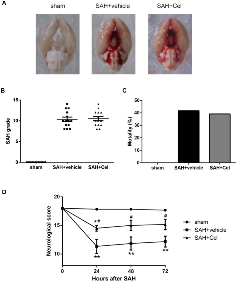
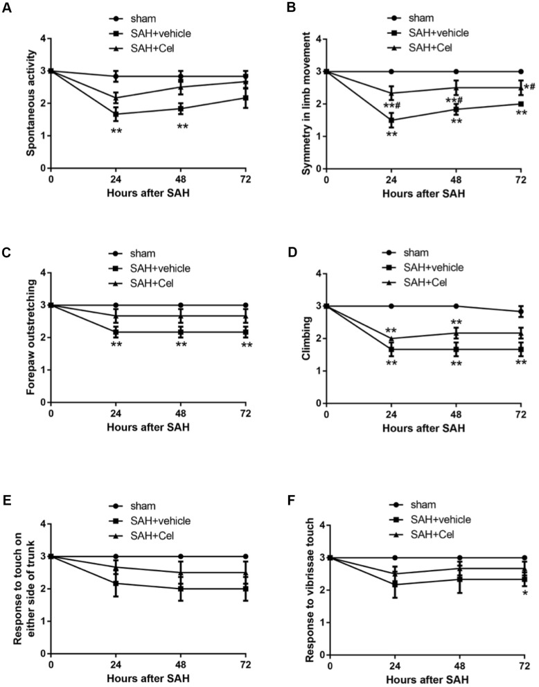
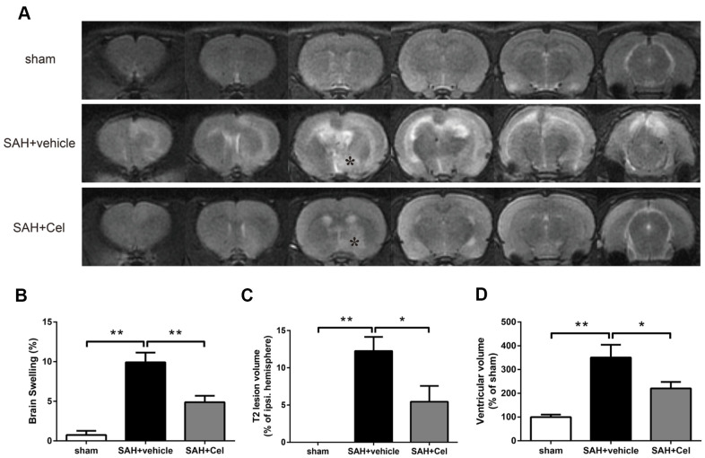
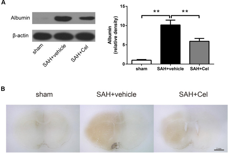
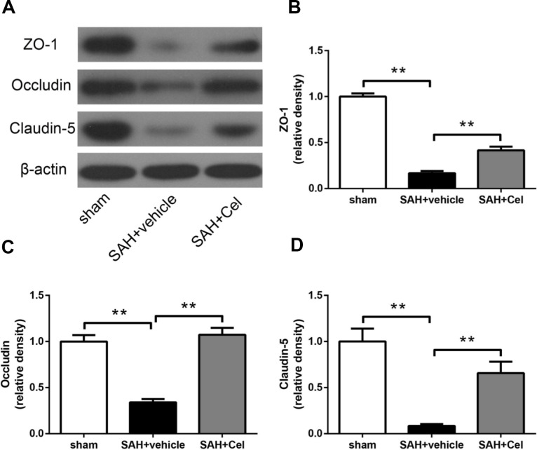
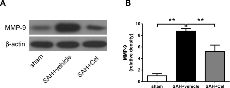
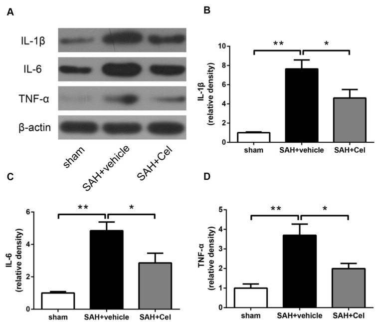
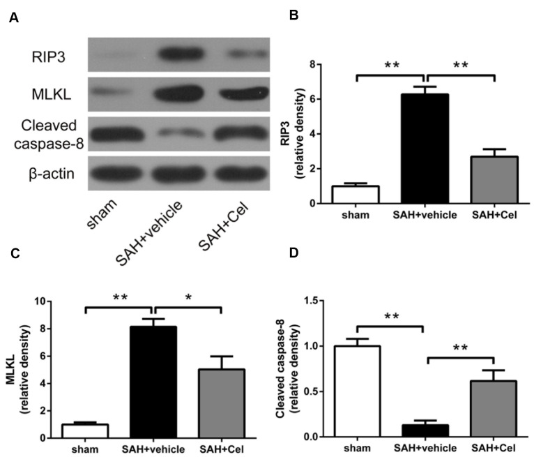
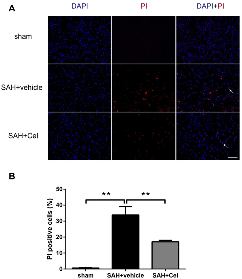
Results: Celastrol treatment attenuated SAH-caused brain swelling, reduced T2 lesion volume and ventricular volume in MRI scanning, and improved overall neurological score. Albumin leakage and the degradation of tight junction proteins were also ameliorated after celastrol administration. Celastrol protected blood-brain bairrer integrity through inhibiting MMP-9 expression and anti-neuroinflammatory effects. Additionally, necroptosis-related proteins RIP3 and MLKL were down-regulated and PI-positive cells in the basal cortex were less in the celastrol-treated SAH group than that in untreated SAH group.

Conclusions: Celastrol exhibits neuroprotective effects on EBI after SAH and deserves to be further investigated as an add-on pharmaceutical therapy.

Keywords: blood-brain barrier; celastrol; early brain injury; necroptosis; subarachnoid hemorrhage.


Conflict of interest statement

CONFLICTS OF INTEREST: The authors declare that they have no conflicts of interest.

Figures

Figure 1
Figure 1
Representative pictures of brains, SAH grade, mortalities and neurological scores at 72 h after SAH. (A) Typical brains of sham, SAH + vehicle, and SAH + Cel group. (B) The grade of SAH severity. (C) SAH-caused mortality rate. (D) Neurological scores at 24, 48 and 72 h after SAH induction. Data were presented as mean±SEM. n = 14. *P < 0.05 versus sham, **P < 0.01 versus sham, #P < 0.05 versus SAH + vehicle.
Figure 2
Figure 2
Neurological subscores at 24, 48 and 72h after SAH induction. (AF) Subscores of (A) spontaneous activity, (B) symmetry in limb movement, (C) forepaw outstretching, (D) climbing, (E) response to touch on either side of trunk, (F) response to vibrissae at each time point after SAH. Data were presented as mean±SEM. n = 14. *P < 0.05 versus sham, **P < 0.01 versus sham, #P < 0.05 versus SAH + vehicle.
Figure 3
Figure 3
Celastrol attenuated brain swelling, reduced T2 lesion volume and ventricular volume after SAH. (A) Representative T2-weighted MRI images (3.0T) of the brains of sham, SAH + vehicle, and SAH + Cel group. (B) Brain swelling was calculated as: ((volume of ipsilateral hemisphere - volume of contralateral hemisphere)/volume of contralateral hemisphere) × 100%. (C) T2 lesion volume was presented as the volume ratio to the ipsilateral hemisphere. (D) Ventricular volume was calculated as Σ(An + An + 1) × d / 2, and was presented as the volume ratio to the average volume of the sham group. Data were presented as mean±SEM. n = 6. *P < 0.05, **P < 0.01.
Figure 4
Figure 4
Celastrol decreased albumin leakage after SAH. (A) Protein levels of albumin in the ipsilateral basal cortex in sham, SAH + vehicle, and SAH + Cel groups at 72 h after SAH induction, detected by WB. (B) Representative histological slides of the albumin staining in the perivascular regions of the ipsilateral basal cortex in sham, SAH + vehicle, and SAH + Cel group. Data were presented as mean±SEM. n = 6. **P < 0.01. Scale bar = 2 mm.
Figure 5
Figure 5
Effects of celastrol treatment on tight junction proteins at 72 h after SAH induction. (A) Representative western blots showing levels of ZO-1, occludin and claudin-5 in the ipsilateral cortex at 72 h after SAH induction. (BD) Quantification of band densities of ZO-1, occludin and claudin-5. The densities of the protein bands were analyzed and normalized to β-actin, and compared to the mean value of the sham group. Data were presented as mean±SEM. n = 6. **P < 0.01.
Figure 6
Figure 6
Effect of celastrol treatment on MMP-9 expression at 72 h after SAH induction. (A) Representative WB showing levels of MMP-9 in the ipsilateral cortex of each group at 72 h after SAH induction. (B) Quantifications of band densities of MMP-9. The densities of the protein bands were analyzed and normalized to β-actin, and compared to the mean value of the sham group. Data were presented as mean±SEM. n = 6. **P < 0.01.
Figure 7
Figure 7
Celastrol decreased neuroinflammation after SAH induction. (A) Representative WB of protein levels of IL-1β, IL-6 and TNF-α in the ipsilateral cortex in each group at 72 h after SAH induction. (BD) The relative band densities of IL-1β, IL-6 and TNF-α. The densities of the protein bands were analyzed and normalized to β-actin, and compared to the mean value of the sham group. Data were presented as mean±SEM. n = 6. *P < 0.05, **P < 0.01.
Figure 8
Figure 8
Celastrol down-regulated RIP3/MLKL signaling pathway after SAH induction. (A) Representative WB showing protein levels of RIP3, MLKL and cleaved caspase-8 in the ipsilateral cortex in each group at 72 h after SAH induction. (BD) Protein quantification of RIP3, MLKL and cleaved caspase-8. The densities of the protein bands were analyzed and normalized to β-actin, and compared to the mean value of the sham group. Data were presented as mean±SEM. n = 6. *P < 0.05, **P < 0.01.
Figure 9
Figure 9
Effects of celastrol on cell injury in the ipsilateral basal cortex at 72 h after SAH induction. (A) Representative microphotographs showed the co-localization of DAPI (blue) with PI (red) positive cells in the ipsilateral basal cortex at 72 h after SAH induction. (B) Quantitative analysis of PI positive cells at 72 h after SAH induction. Data were presented as mean±SEM. n = 6. **P < 0.01. Scale bar = 100 μm.

References

    1. Connolly ES Jr, Rabinstein AA, Carhuapoma JR, Derdeyn CP, Dion J, Higashida RT, Hoh BL, Kirkness CJ, Naidech AM, Ogilvy CS, Patel AB, Thompson BG, Vespa P, and American Heart Association Stroke Council, and Council on Cardiovascular Radiology and Intervention, and Council on Cardiovascular Nursing, and Council on Cardiovascular Surgery and Anesthesia, and Council on Clinical Cardiology. Guidelines for the management of aneurysmal subarachnoid hemorrhage: a guideline for healthcare professionals from the American Heart Association/American Stroke Association. Stroke. 2012; 43:1711–37. 10.1161/STR.0b013e3182587839 - DOI - PubMed
    1. Long B, Koyfman A, Runyon MS. Subarachnoid Hemorrhage: Updates in Diagnosis and Management. Emerg Med Clin North Am. 2017; 35:803–24. 10.1016/j.emc.2017.07.001 - DOI - PubMed
    1. Fujii M, Yan J, Rolland WB, Soejima Y, Caner B, Zhang JH. Early brain injury, an evolving frontier in subarachnoid hemorrhage research. Transl Stroke Res. 2013; 4:432–46. 10.1007/s12975-013-0257-2 - DOI - PMC - PubMed
    1. Cahill J, Zhang JH. Subarachnoid hemorrhage: is it time for a new direction? Stroke. 2009; 40:S86–87. 10.1161/STROKEAHA.108.533315 - DOI - PMC - PubMed
    1. Zolnourian A, Galea I, Bulters D. Neuroprotective Role of the Nrf2 Pathway in Subarachnoid Haemorrhage and Its Therapeutic Potential. Oxid Med Cell Longev. 2019; 2019:6218239. 10.1155/2019/6218239 - DOI - PMC - PubMed

Publication types

MeSH terms