Observational Study
. 2021 Jun 28;22(1):186.
doi: 10.1186/s12931-021-01774-0.

Bronchial thermoplasty in asthma: an exploratory histopathological evaluation in distinct asthma endotypes/phenotypes

Affiliations
Observational Study

Bronchial thermoplasty in asthma: an exploratory histopathological evaluation in distinct asthma endotypes/phenotypes

Eleni Papakonstantinou et al. Respir Res. .

Abstract

Background: Bronchial thermoplasty regulates structural abnormalities involved in airway narrowing in asthma. In the present study we aimed to investigate the effect of bronchial thermoplasty on histopathological bronchial structures in distinct asthma endotypes/phenotypes.

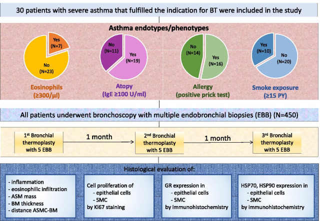
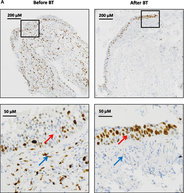
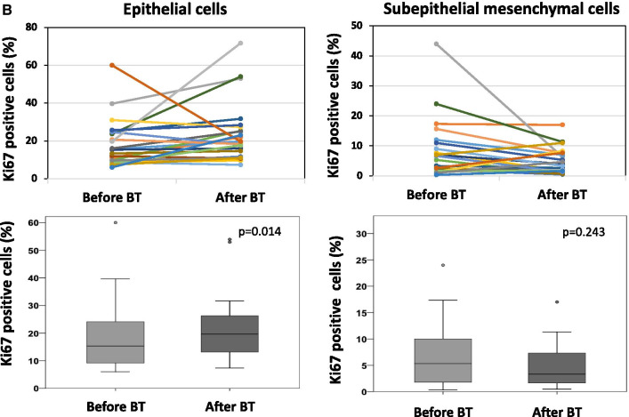
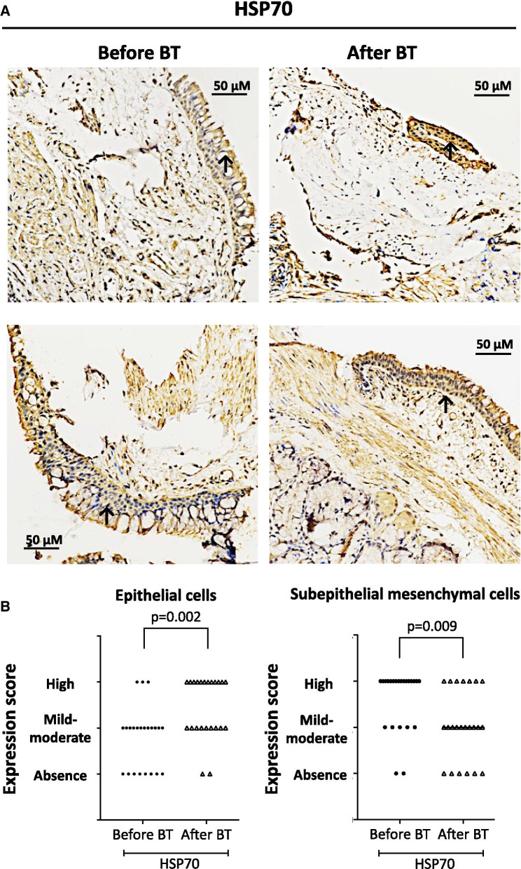
Methods: Endobronchial biopsies (n = 450) were collected from 30 patients with severe uncontrolled asthma before bronchial thermoplasty and after 3 sequential bronchial thermoplasties. Patients were classified based on blood eosinophils, atopy, allergy and smoke exposure. Tissue sections were assessed for histopathological parameters and expression of heat-shock proteins and glucocorticoid receptor. Proliferating cells were determined by Ki67-staining.

Results: In all patients, bronchial thermoplasty improved asthma control (p < 0.001), reduced airway smooth muscle (p = 0.014) and increased proliferative (Ki67 +) epithelial cells (p = 0.014). After bronchial thermoplasty, airway smooth muscle decreased predominantly in patients with T2 high asthma endotype. Epithelial cell proliferation was increased after bronchial thermoplasty in patients with low blood eosinophils (p = 0.016), patients with no allergy (p = 0.028) and patients without smoke exposure (p = 0.034). In all patients, bronchial thermoplasty increased the expression of glucocorticoid receptor in epithelial cells (p = 0.018) and subepithelial mesenchymal cells (p = 0.033) and the translocation of glucocorticoid receptor in the nucleus (p = 0.036). Furthermore, bronchial thermoplasty increased the expression of heat shock protein-70 (p = 0.002) and heat shock protein-90 (p = 0.001) in epithelial cells and decreased the expression of heat shock protein-70 (p = 0.009) and heat shock protein-90 (p = 0.002) in subepithelial mesenchymal cells. The effect of bronchial thermoplasty on the expression of heat shock proteins -70 and -90 was distinctive across different asthma endotypes/phenotypes.

Conclusions: Bronchial thermoplasty leads to a diminishment of airway smooth muscle, to epithelial cell regeneration, increased expression and activation of glucocorticoid receptor in the airways and increased expression of heat shock proteins in the epithelium. Histopathological effects appear to be distinct in different endotypes/phenotypes indicating that the beneficial effects of bronchial thermoplasty are achieved by diverse molecular targets associated with asthma endotypes/phenotypes.

Keywords: Airway smooth muscle; Asthma endotypes; Asthma phenotypes; Bronchial thermoplasty; Epithelial cell regeneration; Glucocorticoid receptor; Heat shock proteins; Severe asthma.


Conflict of interest statement

Dr. Stolz reports grants from Astra-Zeneca AG, Curetis AG, Boston Scientific, Novartis AG, GSK AG, Roche AG, Zambon, Pfizer, Schwabe Pharma AG, Vifor AG. The other authors declare that they have no competing interests related to the study.

Figures

Fig. 1
Fig. 1
The CONSORT flow diagram of the study. All 30 asthma patients fulfilled the criteria for bronchial thermoplasty as they all had persistent, poorly-controlled symptoms, recurrent exacerbations, emergency department visits and hospitalizations despite maximal medical treatment. PY pack years, BM basement membrane, ASM airway smooth muscle, ASMC airway smooth muscle cells, SMC subepithelial mesenchymal cells, EBB endobronchial biopsies
Fig. 2
Fig. 2
Venn diagram that depicts overlaps between different endotypes/phenotypes. Patients with relevant smoke exposure: N = 20; patients with allergy: N = 16; patients with atopy: N = 19; patients with eosinophils ≥ 300 μl: N = 7
Fig. 3
Fig. 3
Proliferative epithelial cells and subepithelial mesenchymal cells in endobronchial biopsies (EBB) obtained before and after BT was assessed by staining with antibodies for Ki67, a nuclear protein that is expressed only in proliferating cells. A Representative microphotographs showing epithelial cells (red arrows) and subepithelial mesenchymal cells (blue arrows) stained positive for Ki67 (brown color). Photographs were captured by Olympus IX83 microscope, using a DS-Ri2 color imaging camera. Right panels show enlargement of the areas in black boxes. B Counting of proliferative cells that stained positive for Ki67 was performed in 3 randomly selected areas on each EBB under the × 200 magnification of the microscope (Nikon Eclipse Ti2 inverted microscope system). Results were expressed as number of Ki67 positive cells per 100 nuclei (%). Upper panels show paired data (from the same patient) before and after BT. In lower panels, horizontal lines in box plots represent median values. Comparisons were made by the Wilcoxon signed rank test. BT bronchial thermoplasty
Fig. 3
Fig. 3
Proliferative epithelial cells and subepithelial mesenchymal cells in endobronchial biopsies (EBB) obtained before and after BT was assessed by staining with antibodies for Ki67, a nuclear protein that is expressed only in proliferating cells. A Representative microphotographs showing epithelial cells (red arrows) and subepithelial mesenchymal cells (blue arrows) stained positive for Ki67 (brown color). Photographs were captured by Olympus IX83 microscope, using a DS-Ri2 color imaging camera. Right panels show enlargement of the areas in black boxes. B Counting of proliferative cells that stained positive for Ki67 was performed in 3 randomly selected areas on each EBB under the × 200 magnification of the microscope (Nikon Eclipse Ti2 inverted microscope system). Results were expressed as number of Ki67 positive cells per 100 nuclei (%). Upper panels show paired data (from the same patient) before and after BT. In lower panels, horizontal lines in box plots represent median values. Comparisons were made by the Wilcoxon signed rank test. BT bronchial thermoplasty
Fig. 4
Fig. 4
Expression of glucocorticoid receptor (GR) in endobronchial biopsies obtained from severe asthma patients using a specific monoclonal antibody. A Representative microphotographs. Lower panels show enlargement of the areas in red boxes. B Representative microphotographs showing increased localization of GR in the nuclei after BT. C expression score in epithelial cells, subepithelial mesenchymal cells and in the nuclei of all cells from patients with severe asthma before and after thermoplasty. The p value was calculated using mixed multinomial (ordinal) logistic regression models, where the factor patient was included as a random effect. BT bronchial thermoplasty
Fig. 5
Fig. 5
Expression of heat shock protein 70 (HSP70) in endobronchial biopsies obtained from severe asthma patients using a specific monoclonal antibody. A Representative microphotographs showing the expression of HSP70 before and after bronchial thermoplasty (BT). B HSP70 expression score in epithelial cells and subepithelial mesenchymal cells from 30 asthma patients before and after BT. The p value was calculated using mixed multinomial (ordinal) logistic regression models, where the factor patient was included as a random effect
Fig. 6
Fig. 6
Expression of heat shock protein 90 (HSP90) in endobronchial biopsies obtained from severe asthma patients using a specific monoclonal antibody. A Representative microphotographs showing the expression of HSP90 before and after bronchial thermoplasty (BT). B HSP90 expression score in epithelial cells and subepithelial mesenchymal cells from 30 asthma patients before and after BT. The p value was calculated using mixed multinomial (ordinal) logistic regression models, where the factor patient was included as a random effect

Similar articles

Cited by

References

    1. Kuruvilla ME, Lee FE, Lee GB. Understanding asthma phenotypes, endotypes and mechanisms of disease. Clin Rev Allergy Immunol. 2019;56(2):219–233. doi: 10.1007/s12016-018-8712-1. - DOI - PMC - PubMed
    1. Wilhelm CP, Chipps BE. Bronchial thermoplasty: a review of the evidence. Ann Allergy Asthma Immunol. 2016;116:92–98. doi: 10.1016/j.anai.2015.11.002. - DOI - PubMed
    1. Pavord ID, Cox G, Thomson NC, RISA Trial Study Group et al. Safety and efficacy of bronchial thermoplasty in symptomatic, severe asthma. Am J Respir Crit Care Med. 2007;176:1185–1191. doi: 10.1164/rccm.200704-571OC. - DOI - PubMed
    1. Castro M, Rubin AS, Laviolette M, et al. Effectiveness and safety of bronchial thermoplasty in the treatment of severe asthma. Am J Respir Crit Care Med. 2010;181:116–124. doi: 10.1164/rccm.200903-0354OC. - DOI - PMC - PubMed
    1. Chernyavsky IL, Russell RJ, Saunders RM, et al. In vitro, in silico and in vivo study challenges the impact of bronchial thermoplasty on acute airway smooth muscle mass loss. Eur Respir J. 2018;51:1701680. doi: 10.1183/13993003.01680-2017. - DOI - PMC - PubMed

Publication types