. 2021 Jul 13;118(28):e2101823118.
doi: 10.1073/pnas.2101823118.

Neural specification, targeting, and circuit formation during visual system assembly

Affiliations

Neural specification, targeting, and circuit formation during visual system assembly

Jennifer Malin et al. Proc Natl Acad Sci U S A. .

Abstract

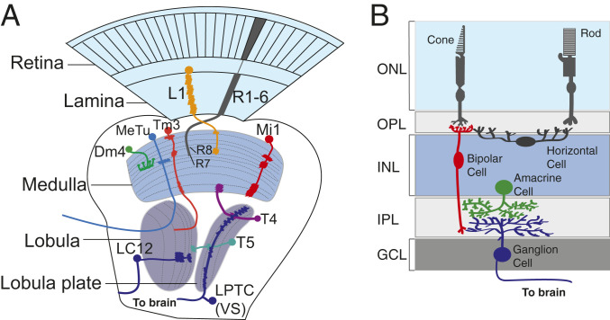
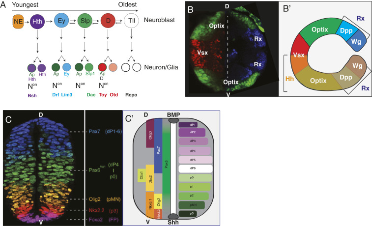
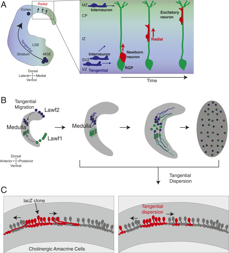
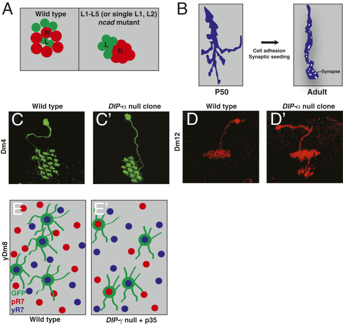
Like other sensory systems, the visual system is topographically organized: Its sensory neurons, the photoreceptors, and their targets maintain point-to-point correspondence in physical space, forming a retinotopic map. The iterative wiring of circuits in the visual system conveniently facilitates the study of its development. Over the past few decades, experiments in Drosophila have shed light on the principles that guide the specification and connectivity of visual system neurons. In this review, we describe the main findings unearthed by the study of the Drosophila visual system and compare them with similar events in mammals. We focus on how temporal and spatial patterning generates diverse cell types, how guidance molecules distribute the axons and dendrites of neurons within the correct target regions, how vertebrates and invertebrates generate their retinotopic map, and the molecules and mechanisms required for neuronal migration. We suggest that basic principles used to wire the fly visual system are broadly applicable to other systems and highlight its importance as a model to study nervous system development.

Keywords: Drosophila; neural development; patterning; retina; visual system.


Conflict of interest statement

The authors declare no competing interest.

Figures

Fig. 1.
Fig. 1.
Drosophila and mammalian visual system organization. (A) Visual input in Drosophila is captured by photoreceptors divided into ∼800 ommatidia. Outer photoreceptor axons (R1 to R6) project to the cartridges of the lamina, while inner photoreceptor axons (R7 to R8) project to the medulla. Lamina neurons (e.g., L1) also project their axons to the medulla (orange–yellow). Medulla neurons can be divided into numerous classes. Mi neurons (Mi1; red) project their arbors throughout the entire medulla. Transmedullary neurons (e.g., Tm3; orange) connect the medulla to the lobula. Distal medulla (e.g., Dm4; green) neurons are multicolumnar and project arbors across multiple medulla columns. The lobula and lobula plate neuropils are responsible for processing different aspects of vision. T4 neurons (purple) connect the lobula plate to the proximal medulla, while T5 neurons (teal) connect the lobula/lobula plate, which processes broad field motion. LC (e.g., LC12) neurons (dark blue) project within the lobula and send an arbor to the central brain to process various visual features. LPTCs (e.g., VS neurons) are sensitive to wide-field motion and project their arbors to the central brain, as do medulla tubercule (bright blue) neurons. (B) Input to the mammalian visual system is captured by photoreceptors, which are categorized as dim light–sensing rods or bright light/color–sensing cones. Rod and cone photoreceptors synapse onto rod or cone bipolar cells (red), respectively. Horizontal cells (dark gray) integrate the input from multiple photoreceptor cells to bipolar cells. Bipolar cells (red) make synapses with feature-detecting RGCs (blue). RGCs project neurites to higher-order processing centers. Amacrine cells (green) modulate bipolar to RGC signaling. Like Dm neurons in the fly, they are broadly arborizing. Müller glia are integral to visual system processing but are not shown in the figure. GCL, ganglion cell layer; INL, inner nuclear layer; IPL, inner plexiform layer; ONL, outer nuclear layer; OPL, outer plexiform layer.
Fig. 2.
Fig. 2.
Spatiotemporal patterning/Notch signaling provide neuronal diversity in the visual system. (A) Drosophila OPC medulla neuroblasts express transcription factors in a temporal cascade whose output defines the identities of neurons born from those cells. Notchon/off signaling in the daughters of dividing Ganglion Mother Cells further diversifies neuronal fate. (B, B′) The OPC neuroepithelium can be subdivided into multiple regions based on the expression of transcription factors: Rx (blue) is expressed at the tips, Optix (red) is expressed in the middle, and Vsx (green) is expressed in the center. Image was generated by splicing two images (one of Vsx and Optix and the other with Optix and Rx) together. Growth factor Hh (B′, orange) is expressed in the ventral OPC, whereas the Rx domain can be subdivided into Wg and Dpp expressing regions. Dotted line: splice site. (C) Immunofluorescence image of spatially regulated transcription factor (TF) expression, which regionalizes progenitors to allow for the generation of different classes of spinal cord neurons. (Adapted with permission from ref. .) (C′) Bone morphogenetic protein (BMP) morphogen signaling activates the expression of basic helix-loop-helix (bHLH) TFs such as Olig3 (violet) to pattern the six classes of neurons in the dorsal spinal cord. Shh signaling suppresses the expression of Class I homeodomain TFs (e.g., Pax7, Dbx1, Dbx2, and Pax6) while activating the expression of Class II factors (e.g., Nkx2.2). The intersection of these gene expression patterns demarcates five progenitor classes in the ventral spinal cord.
Fig. 3.
Fig. 3.
Cell migration is a conserved phenomenon utilized across numerous neural types and many species. (A) Inhibitory interneurons born in the Medial Ganglionic Eminence (MGE) of the ventral telencephalon migrate tangentially into the cortical wall before dispersing in the cortical plate. (Inset) Excitatory cortical neurons are born from RGPs and radially migrate along glial fibers to reach their appropriate cortical layer. (B) Like inhibitory interneurons, developing Lawf neurons in Drosophila migrate tangentially to reach the medulla cortex. Later, Lawf1 and Lawf2 tangentially disperse across the medulla to reach their final positions. (C) Mosaic clones of ChAT-expressing mouse retinal amacrine cells (red) tangentially disperse away from their siblings (Right) to find their final positions; as a result, they are found intermingled among transgene-negative (gray) amacrine cells (186). Adapted from ref. , with permission from Elsevier. ChAT, choline acetyltransferase; CP, cortical plate; IZ: intermediate zone; LGE, lateral ganglionic eminence; MZ, marginal zone; SVZ, subventricular zone; VZ, ventricular zone.
Fig. 4.
Fig. 4.
Cell adhesion molecules promote different aspects of nervous system assembly. (A) In the Drosophila lamina, differential adhesion promoted by high levels of NCad (lamina neurons [L]; green) or lower levels (photoreceptors [R]; red) directs lamina and neurons to their appropriate positions within a cartridge, internal for lamina neurons, periphery for photoreceptors. NCad mutants (Right) push lamina neurons from the cartridge center to the periphery. (B) Filopodial extensions from Drosophila photoreceptor axons are highly dynamic and explore their surroundings during pupation (Left). Later, cell adhesion molecules and synaptic components exponentially increase the stability of the axon, leading to column refinement and synaptogenesis (Right). (CE) IgSF-containing DIPs/Dprs promote synaptogenesis by promoting adhesion between synaptic partners. DIP-mutant neurites target fewer columns (C and C′), target inappropriate sublayers (D and D′), or target the incorrect partners (E and E′). C, C', D, and D' are reprinted from ref. , with permission from Elsevier. E and E' are adapted from ref. , with permission from AAAS. GFP, green fluorescent protein; P50, 50% pupariation; pR7, pale photoreceptor R7; yR7, yellow photoreceptor R7.

References

    1. Luo L., Flanagan J. G., Development of continuous and discrete neural maps. Neuron 56, 284–300 (2007). - PubMed
    1. Fischbach K.-F., Dittrich A. P. M., The optic lobe of Drosophila melanogaster. I. A Golgi analysis of wild-type structure. Cell Tissue Res. 258, 441–475 (1989).
    1. Jenett A., et al., A GAL4-driver line resource for Drosophila neurobiology. Cell Rep. 2, 991–1001 (2012). - PMC - PubMed
    1. Pfeiffer B. D., et al., Tools for neuroanatomy and neurogenetics in Drosophila. Proc. Natl. Acad. Sci. U.S.A. 105, 9715–9720 (2008). - PMC - PubMed
    1. Macosko E. Z., et al., Highly parallel genome-wide expression profiling of individual cells using nanoliter droplets. Cell 161, 1202–1214 (2015). - PMC - PubMed

Publication types

LinkOut - more resources