. 2021 Jun 28;11(1):13371.
doi: 10.1038/s41598-021-92650-w.

Accelerated burn wound healing with photobiomodulation therapy involves activation of endogenous latent TGF-β1

Affiliations

Accelerated burn wound healing with photobiomodulation therapy involves activation of endogenous latent TGF-β1

Imran Khan et al. Sci Rep. .

Erratum in

Abstract

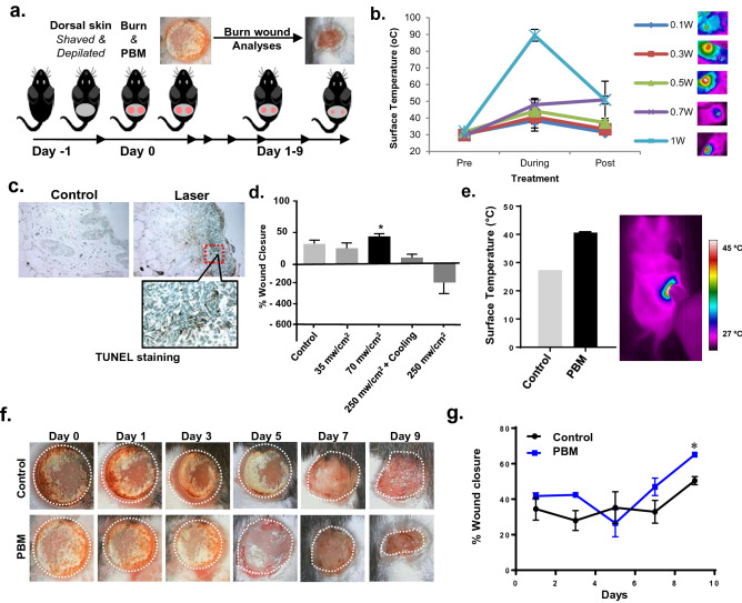
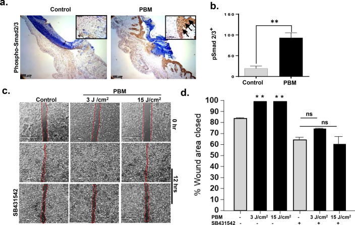
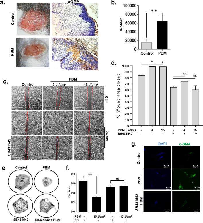
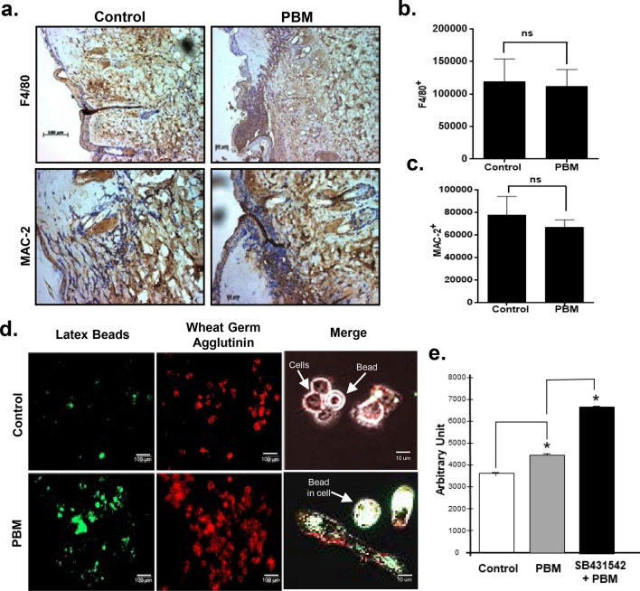
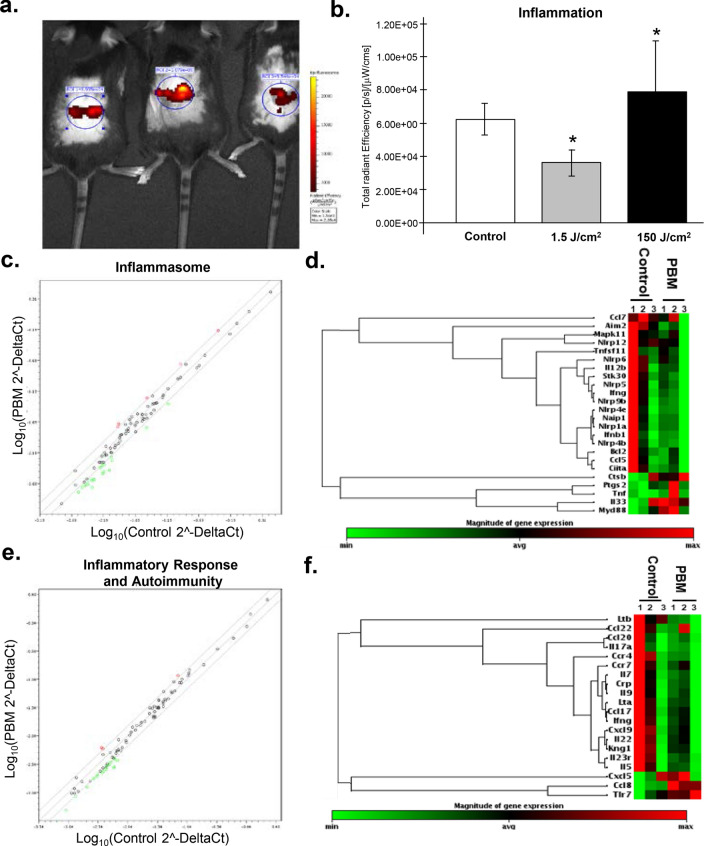
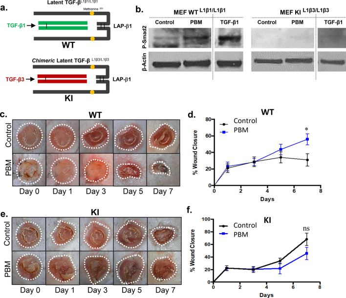
The severity of tissue injury in burn wounds from associated inflammatory and immune sequelae presents a significant clinical management challenge. Among various biophysical wound management approaches, low dose biophotonics treatments, termed Photobiomodulation (PBM) therapy, has gained recent attention. One of the PBM molecular mechanisms of PBM treatments involves photoactivation of latent TGF-β1 that is capable of promoting tissue healing and regeneration. This work examined the efficacy of PBM treatments in a full-thickness burn wound healing in C57BL/6 mice. We first optimized the PBM protocol by monitoring tissue surface temperature and histology. We noted this dynamic irradiance surface temperature-monitored PBM protocol improved burn wound healing in mice with elevated TGF-β signaling (phospho-Smad2) and reduced inflammation-associated gene expression. Next, we investigated the roles of individual cell types involved in burn wound healing following PBM treatments and noted discrete effects on epithelieum, fibroblasts, and macrophage functions. These responses appear to be mediated via both TGF-β dependent and independent signaling pathways. Finally, to investigate specific contributions of TGF-β1 signaling in these PBM-burn wound healing, we utilized a chimeric TGF-β1/β3 knock-in (TGF-β1Lβ3/Lβ3) mice. PBM treatments failed to activate the chimeric TGF-β1Lβ3/Lβ3 complex and failed to improve burn wound healing in these mice. These results suggest activation of endogenous latent TGF-β1 following PBM treatments plays a key role in burn wound healing. These mechanistic insights can improve the safety and efficacy of clinical translation of PBM treatments for tissue healing and regeneration.


Conflict of interest statement

The authors declare no competing interests.

Figures

Figure 1
Figure 1
PBM treatment improves burn wound healing. (a) Representative diagram showing scheme of wound healing experiment. On the 1st day, the dorsal skin of 5-week-old C57BL/6NCr male mice was shaved, and on the 2nd day, two burn wounds were created, followed by PBM treatments (Day 0) with an 810 nm CW diode laser. Burn wounds were photographed every other day up to day 9; (b) Tissue surface temperature was monitored during laser treatments with increasing irradiances with a thermal camera; (c) Tissue damage was assessed in these tissues with TUNEL staining indicating phototoxicity at skin temperature above 45 °C; (d) Burn wound healing following PBM treatments at increasing doses and concomitant surface cooling, results are expressed as means and SDs representative of two independent experiments, significance was determined using non-parametric Student's t-test with *p-value < 0.05; (e) Optimal PBM dose treatments ensured skin surface temperature was > 45 °C using a dynamic irradiance protocol assessed with thermal imaging (left) and quantitation (right); (f) PBM treatments on burn wounds were photographed every other day for up to 9 days and compared to untreated controls; (g) Wound areas were digitally quantitated, results are expressed as means and SDs that is representative of five independent experiments, significance was determined using non-parametric Student's T-test with n = 8, *p-value < 0.05.
Figure 2
Figure 2
PBM activated TGF-β signaling promotes burn wound epithelial migration. (a) Burn wound tissues were immunostained for p-Smad2 at day 9; (b) Digital quantitation of immunohistochemical staining from mice sections, means and SDs are shown (n = 8, *p < 0.05, unpaired Student's T-Test); (c) Human dermal keratinocytes, HaCaT cells were plated in a 6-well tissue culture plate and were allowed to form confluent cultures for 24 h, and a scratch wound was created. PBM treatments at different doses with or without SB431542 inhibitor was performed, and images were captured with a digital microscope at 12 h; (d) Images were quantitated using T scratch software, and % area closed are shown as means and SDs that is representative of two independent experiments performed with replicates, significance was determined using one-way ANOVA among different treatments using the Tukey's multiple comparisons test indicated as **p < 0.005, *p < 0.05 and n.s. not significant.
Figure 3
Figure 3
PBM activated myofibroblasts in burn wounds promotes wound contraction. (a) PBM treated mice were sacrificed, and wound areas were immunostained for α-SMA on day 9; (b) Digital quantitation of immunohistochemical staining from mice sections, means and SDs are shown (n = 8, unpaired T-Test, **p < 0.005); (c) Human dermal fibroblast cells were plated in a 6-well tissue culture plate and were allowed to form confluent cultures for 24 h, and a scratch wound was created. PBM treatments at different doses with or without SB431542 inhibitor was performed, and images were captured with a digital microscope at 12 h; (d) Images were quantitated using T scratch software, and % area closed are shown as means and SDs that is representative of two independent experiments performed with replicates, significance was determined using one-way ANOVA among different treatments using the Tukey's multiple comparisons test indicated as *p < 0.05 and n.s. not significant; (e) Collagen gel contraction assays were performed with dermal fibroblast cells cast in collagen gels plated in 24-well culture dishes. PBM treatments were performed at various doses with or without prior incubation with SB431542, and gels were then photographed after 24 h; (f) Gel areas were plotted, and data is shown as means and SDs that is representative of two independent experiments performed with replicates, statistical significance was determined with one-way ANOVA among different treatments using the Tukey's multiple comparisons tests, **p < 0.005; (g) Gels were fixed and immunostained for αSMA, and representative fluorescence images are shown.
Figure 4
Figure 4
PBM treatments increased macrophage phagocytic activity. (a) Burn wound tissues from untreated and PBM treated mice at 24 h post-injury were immunostained for the macrophage markers F4/80 and Mac-2; (b) Quantitation of F4/80 immunohistochemical staining from mice sections is represented as means and SDs (n = 8, unpaired Student's T-Test, n.s. not significant); (c) Quantitation of Mac-2 immunohistochemical staining is shown as means and SDs are shown (n = 8, unpaired Student's T-Test, n.s. not significant); (d) Macrophage cell line RAW 264.7 cells were seeded in 96 well plates and incubated with FITC-labelled latex beads to examine phagocytosis. Cell membranes were counterstained with Texas Red-conjugated Wheat Germ Agglutinin and imaged using a fluorescence microscope; (e). Fluorescent intensity was quantitated using NIH ImageJ, and data are presented as means and SDs that are representative of two independent experiments performed with replicates; statistical significance was determined with one-way ANOVA among different treatments using the Tukey's multiple comparisons test, *p < 0.05.
Figure 5
Figure 5
PBM downregulates inflammatory response in burn wounds. (a) Laser treatments were performed at varying doses and 24 h later mice were anesthetized and injected with the inflammation detection probe (XenoLight RediJect, 200 mg/kg, 100 μl per mice, i.p.) and live fluorescence imaging was performed; (b) Quantitation of fluorescence images using the Living Image software (Perkin-Elmer) and results are shown as means and SDs representative of two independent experiments with replicates, significance was determined using non-parametric Student's t-test with *p value < 0.05; (c) qPCR arrays were performed on wound tissues at 24 h of post-PBM treatments using inflammasome array and data is shown as a pair-wise comparison of gene expression in control versus PBM treated mice burn wounds with XY-scatterplot analysis of log base 2-transformed expression data, results represent two independent studies performed with triplicates; each dot represents a gene, with red dots showing genes denoting downregulated genes following PBM treatments with an FDR corrected p < 0.05, green dots represents upregulated genes with FDR < 0.05 and black dots are genes whose expression was similar between the two groups; (d) Hierarchical clustering of differentially regulated genes in untreated versus PBM treated burn wounds in the inflammasome array; (e) Similar qPCR arrays were performed on wound tissues at 24 h of post-PBM treatments using the inflammatory response and autoimmunity array and data is similarly presented as described above; (f) Hierarchical clustering of differentially regulated genes in the latter gene expression array.
Figure 6
Figure 6
Lack of PBM-activated TGF-β fails to promote burn wounds in chimeric mice. (a) Schematic for wild type latent TGF-βL1β1/L1β1 and chimeric latent TGF-βL1β3/L1β3 knock-in mouse models; (b) MEFs obtained from both mice were PBM treated, lysed, and assessed for phospho-Smad2 by western blotting, recombinant TGF-β1 treatment was used as a positive control; (c) Burn wound healing and PBM treatments were performed in the WT mice, and digital images were captured for up to 7 days; (d) Wound areas were digitally quantitated, results are expressed as means and SDs representative of two independent experiments, n = 6, significance was determined using non-parametric Student's t-test with *p-value < 0.05; (c) Similarly, burn wound healing and PBM treatments were performed in the KI mice, and digital images were captured for up to 7 days; (d) Wound areas were digitally quantitated, results are expressed as means and SDs representative of two independent experiments, n = 6, significance was determined using non-parametric Student's t-test with, n.s. not significant.

References

    1. Penn JW, Grobbelaar AO, Rolfe KJ. The role of the TGF-beta family in wound healing, burns and scarring: a review. Int. J. Burns Trauma. 2012;2:18–28. - PMC - PubMed
    1. Monstrey S, Hoeksema H, Verbelen J, Pirayesh A, Blondeel P. Assessment of burn depth and burn wound healing potential. Burns. 2008;34:761–769. doi: 10.1016/j.burns.2008.01.009. - DOI - PubMed
    1. Cancio LC, Salinas J, Kramer GC. Protocolized resuscitation of burn patients. Crit Care Clin. 2016;32:599–610. doi: 10.1016/j.ccc.2016.06.008. - DOI - PubMed
    1. Gibran NS, Heimbach DM. Current status of burn wound pathophysiology. Clin. Plast. Surg. 2000;27:11–22. doi: 10.1016/S0094-1298(20)32680-8. - DOI - PubMed
    1. Gurtner GC, Werner S, Barrandon Y, Longaker MT. Wound repair and regeneration. Nature. 2008;453:314–321. doi: 10.1038/nature07039. - DOI - PubMed

Publication types

Substances