. 2021 Jun 28;11(1):13433.
doi: 10.1038/s41598-021-92824-6.

Rspo2 inhibits TCF3 phosphorylation to antagonize Wnt signaling during vertebrate anteroposterior axis specification

Affiliations

Rspo2 inhibits TCF3 phosphorylation to antagonize Wnt signaling during vertebrate anteroposterior axis specification

Alice H Reis et al. Sci Rep. .

Abstract

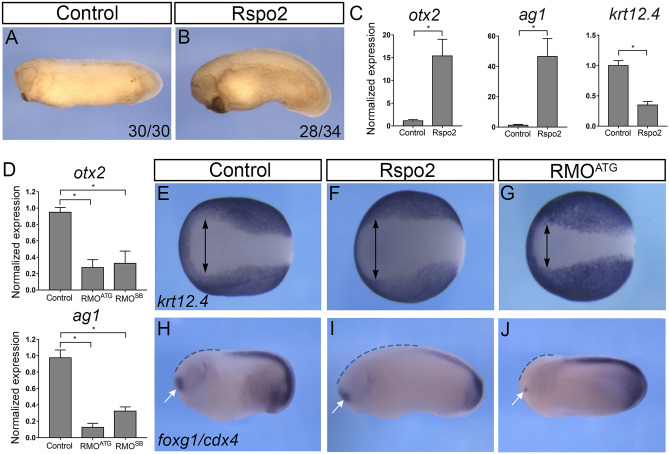
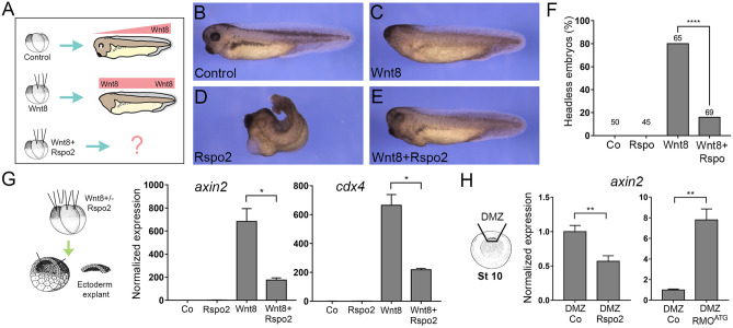
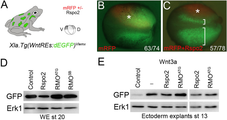
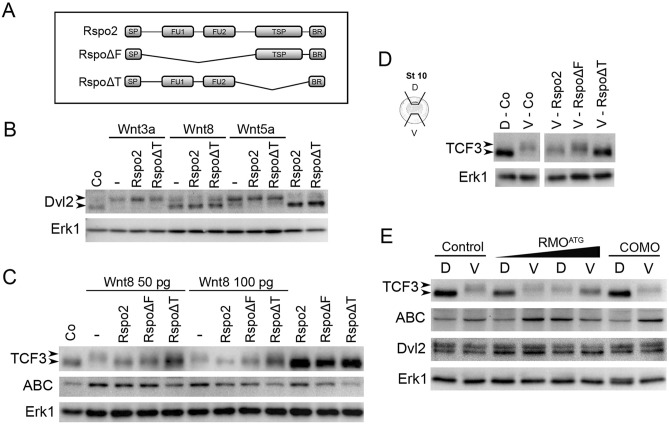
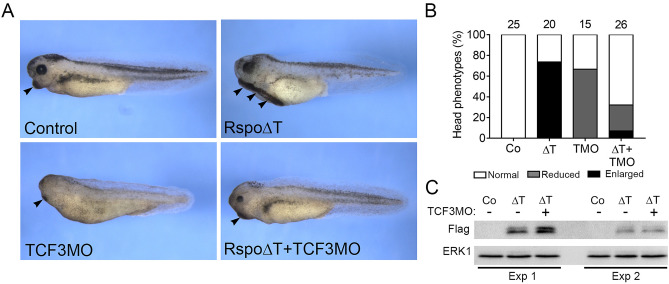
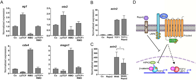
The Wnt pathway activates target genes by controlling the β-catenin-T-cell factor (TCF) transcriptional complex during embryonic development and cancer. This pathway can be potentiated by R-spondins, a family of proteins that bind RNF43/ZNRF3 E3 ubiquitin ligases and LGR4/5 receptors to prevent Frizzled degradation. Here we demonstrate that, during Xenopus anteroposterior axis specification, Rspo2 functions as a Wnt antagonist, both morphologically and at the level of gene targets and pathway mediators. Unexpectedly, the binding to RNF43/ZNRF3 and LGR4/5 was not required for the Wnt inhibitory activity. Moreover, Rspo2 did not influence Dishevelled phosphorylation in response to Wnt ligands, suggesting that Frizzled activity is not affected. Further analysis indicated that the Wnt antagonism is due to the inhibitory effect of Rspo2 on TCF3/TCF7L1 phosphorylation that normally leads to target gene activation. Consistent with this mechanism, Rspo2 anteriorizing activity has been rescued in TCF3-depleted embryos. These observations suggest that Rspo2 is a context-specific regulator of TCF3 phosphorylation and Wnt signaling.


Conflict of interest statement

The authors declare no competing interests.

Figures

Figure 1
Figure 1
Rspo2 function is essential for anterior development. (A,B) Four-cell embryos were injected with 0.5 ng of Rspo2 RNA into both animal-ventral blastomeres and cultured until stage 28. (A) Uninjected control embryo. (B) Representative embryo injected with Rspo2 RNA. The penetrance is indicated as the ratio of the number of embryos with the phenotype and the total number of injected embryos. (C) The effect of Rspo2 on gene marker expression. Animal pole explants were dissected at stage 9 from embryos overexpressing Rspo2 RNA or uninjected controls. RT-qPCR analysis was carried out for otx2, ag1, and krt12.4 at stage 18. (D) Altered gene expression in Rspo2 morphants. RNA was isolated from stage 18 control embryos or embryos depleted of Rspo2. RT-qPCR for ag1 and otx2 was carried out in triplicates. (C,D) Each graph is a single experiment with triplicate samples, representative from at least 3 independent experiments. Means + /− s. d. are shown. Statistical significance has been assessed by Student’s t test, *, p < 0.05. (EJ) In situ hybridization of control and manipulated stage 16 or 25 embryos with krt12.4 (EG), foxg1 and cdx4 (HJ) probes. (EG) Width of the anterior neural plate is shown as lack of krt12.4 (arrows). (HJ) The foxg1 domain is indicated by white arrows, the anterior region lacking cdx4—by dashed lines. See Supplementary Table 1 for quantification.
Figure 2
Figure 2
Rspo2 antagonizes Wnt signaling. (A) Scheme of the experiment. Four-cell embryos were injected animally into both dorsal blastomeres with the indicated constructs and cultured to stage 38. (B) Uninjected control embryo. (C) Headless embryo injected with Wnt8 DNA (50 pg). (D) Embryo injected with Rspo2 RNA (0.5 ng). (E) Embryo coexpressing Wnt8 DNA and Rspo2 mRNA. (F) Quantification of the data in (AD) representative of 3 independent experiments. Numbers of embryos per group are shown above each bar. ****, p < 0.0001, Fisher's exact test. (G) Target gene expression in Wnt8 and Rspo2-stimulated ectoderm explants. Embryos were injected into four animal blastomeres with Wnt8 DNA (50 pg) and Rspo2 RNA (0.5 ng), as indicated, and ectoderm explants were prepared at stage 9 and cultured until stage 13. (H) Dorsal marginal zones (DMZ) were dissected at stage 10 from the control and Rspo2 RNA- or RMOATG-injected embryos and cultured until stage 12. (G,H) RT-qPCR analysis was carried out in triplicates for axin2 and cdx4, and normalized to eef1a1 levels. Means + /− s.d. are shown. Graphs are representative of three independent experiments. Statistical significance has been assessed by Student’s t test, *, p < 0.05; **, p < 0.01.
Figure 3
Figure 3
The effects of Rspo2 manipulation on Wnt reporter activity in transgenic embryos. (A) Experimental scheme. Xla.Tg(WntREs:dEGFP)Vlemx embryos were injected into one dorsal blastomere with mRFP RNA (50 pg) with (C) or without (B) Rspo2 RNA (0.5 ng). GFP fluorescence of the injected embryos at stage 18 is shown. Embryo images are representative of 3 different experiments. Asterisk indicates the injected side of the embryo, brackets in C show the comparison of the injected and the control sides. (D,E) Rspo2 modulates Wnt reporter activation. (D) Rspo2 RNA (0.5 ng), RMOATG (10 ng) or RMOSB (20 ng) were injected at two dorsal blastomeres at 4-cell stage. The embryos were lysed at stage 20 and immunoblotted with anti-GFP antibodies. (E) Four-cell stage embryos were injected animally with Wnt3a RNA (50 pg) and Rspo2 RNAs (0.5 ng) or RMOATG (10 ng). Ectoderm explants were dissected at stage 9 and cultured until stage 13, then lysed and immunoblotted with anti-GFP antibodies. Erk1 is a control for loading. In (E) two right lanes were run in the same gel but away from the left lanes (see Supplementary Fig. 5). Five embryos or 10 explants were pooled for each experimental condition in (D,E).
Figure 4
Figure 4
Rspo2 inhibits TCF3 phosphorylation. (A) Schematic of Rspo2 deletion constructs. SP, signal peptide; FU1, furin-like domain 1; FU2, furin-like domain 2; TSP, thrombospondin type 1 domain; BR, the basic amino acid-rich domain. (B,C) Effects of Rspo2 constructs on Wnt-dependent Dvl2 phosphorylation (B) and TCF3 phosphorylation and β-catenin levels (C). Four-cell stage embryos were injected animally with Wnt8 DNA (50 pg or 100 pg) or Wnt8, Wnt3a or Wnt5a RNAs (1 ng each) and Rspo2, Rspo∆F or Rspo∆T RNAs (0.5 ng each) as indicated. Ectoderm explants were dissected at stage 9 and cultured until stage 12 for immunoblotting with antibodies against Dvl2, TCF3, ABC (non-phosphorylated β-catenin). Arrowheads indicate the position of phosphorylated (upshifted) and non-phosphorylated Dvl2 or TCF3 proteins. Erk1 controls for loading. (D) Effects of Rspo2 constructs (0.5 ng each) on TCF3 phosphorylated by endogenous signals. Dorsal marginal zone (D) and ventral marginal zone (V) were dissected from the control and injected embryos at stage 10 and cultured until stage 12.5 for immunoblotting with anti-TCF3 antibodies as shown. Control D and V groups were run in the same gel but separately from the other groups (see Supplementary Fig. 5). (E) Effects of Rspo2 depletion on TCF3 phosphorylation by endogenous signals. DMZ and VMZ explants of embryos injected with control MO (COMO, 20 ng) or RMOATG (20 ng) were dissected and analyzed by immunoblotting as in (B,C).
Figure 5
Figure 5
TCF3 is essential for Rspo2 inhibitory effects. (A) TCF3MO rescues the anteriorized phenotype of Rspo∆T RNA overexpressing embryos. Four-cell stage embryos were dorsally injected with TCF3MO (30 ng) and/or Rspo∆T RNA (0.5 ng). Arrowheads indicate the cement gland. (B) Quantification of the data in (A) representative of two independent experiments. Numbers of embryos per group are shown above each bar. (C) Rspo∆T expression levels are not altered by TCF3MO in ectoderm explants (stage 12) in two independent experiments (Exp 1 and Exp 2). ∆T, Rspo∆T; TMO, TCF3MO.
Figure 6
Figure 6
Rspo2 inhibits Wnt signaling through TCF3. (A) ∆βTCF3 RNA (10 pg) rescues ag1, otx2, cdx4, and msgn1 expression in embryos injected with 10 ng of RMOATG. B, C, Rspo2 inhibits axin2 upregulation by Wnt8 (C) but not TCF1 (B) in ectoderm cells. Embryos were injected with Wnt8 (20 pg) or TCF1 (100 pg) RNA without or with Rspo2 RNA (300 pg). Ectoderm explants were prepared at stage 8.5–9 and analyzed at stage 13. RT-qPCR analysis was carried out in triplicates for axin2 and normalized to eef1a1 levels. Means + /− s.d. are shown. Graphs are representative of 2–4 independent experiments. Statistical significance has been assessed by Student’s t test, *, p < 0.05. D, Model for Rspo2-mediated repression. Rspo2 functions via an unknown receptor to inhibit Wnt target gene activation mediated by TCF3 phosphorylation but not TCF1-dependent transcriptional responses.

Similar articles

Cited by

References

    1. MacDonald BT, Tamai K, He X. Wnt/beta-catenin signaling: components, mechanisms, and diseases. Dev. Cell. 2009;17:9–26. doi: 10.1016/j.devcel.2009.06.016. - DOI - PMC - PubMed
    1. Nusse R, Clevers H. Wnt/beta-catenin signaling, disease, and emerging therapeutic modalities. Cell. 2017;169:985–999. doi: 10.1016/j.cell.2017.05.016. - DOI - PubMed
    1. Hikasa H, Ezan J, Itoh K, Li X, Klymkowsky MW, Sokol SY. Regulation of TCF3 by Wnt-dependent phosphorylation during vertebrate axis specification. Dev. Cell. 2010;19:521–532. doi: 10.1016/j.devcel.2010.09.005. - DOI - PMC - PubMed
    1. Kim CH, Oda T, Itoh M, Jiang D, Artinger KB, Chandrasekharappa SC, Driever W, Chitnis AB. Repressor activity of Headless/Tcf3 is essential for vertebrate head formation. Nature. 2000;407:913–916. doi: 10.1038/35038097. - DOI - PMC - PubMed
    1. Nguyen H, Rendl M, Fuchs E. Tcf3 governs stem cell features and represses cell fate determination in skin. Cell. 2006;127:171–183. doi: 10.1016/j.cell.2006.07.036. - DOI - PubMed

Publication types

MeSH terms

Substances