. 2021 Aug;18(8):1861-1870.
doi: 10.1038/s41423-021-00720-w. Epub 2021 Jun 28.

Self-activation of Vγ9Vδ2 T cells by exogenous phosphoantigens involves TCR and butyrophilins

Affiliations

Self-activation of Vγ9Vδ2 T cells by exogenous phosphoantigens involves TCR and butyrophilins

Chloé Laplagne et al. Cell Mol Immunol. 2021 Aug.

Abstract

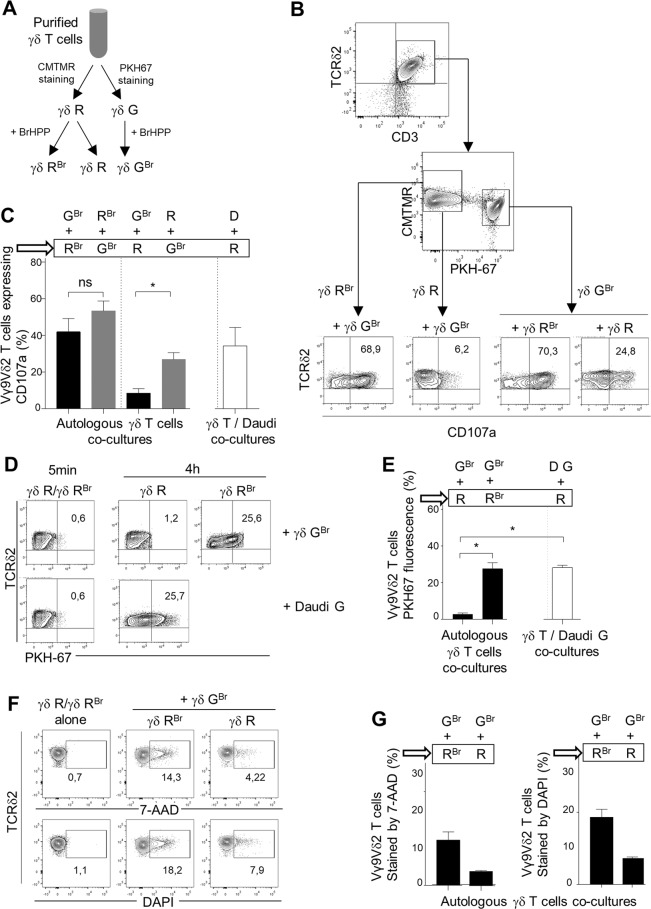
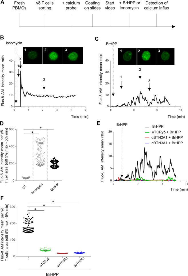
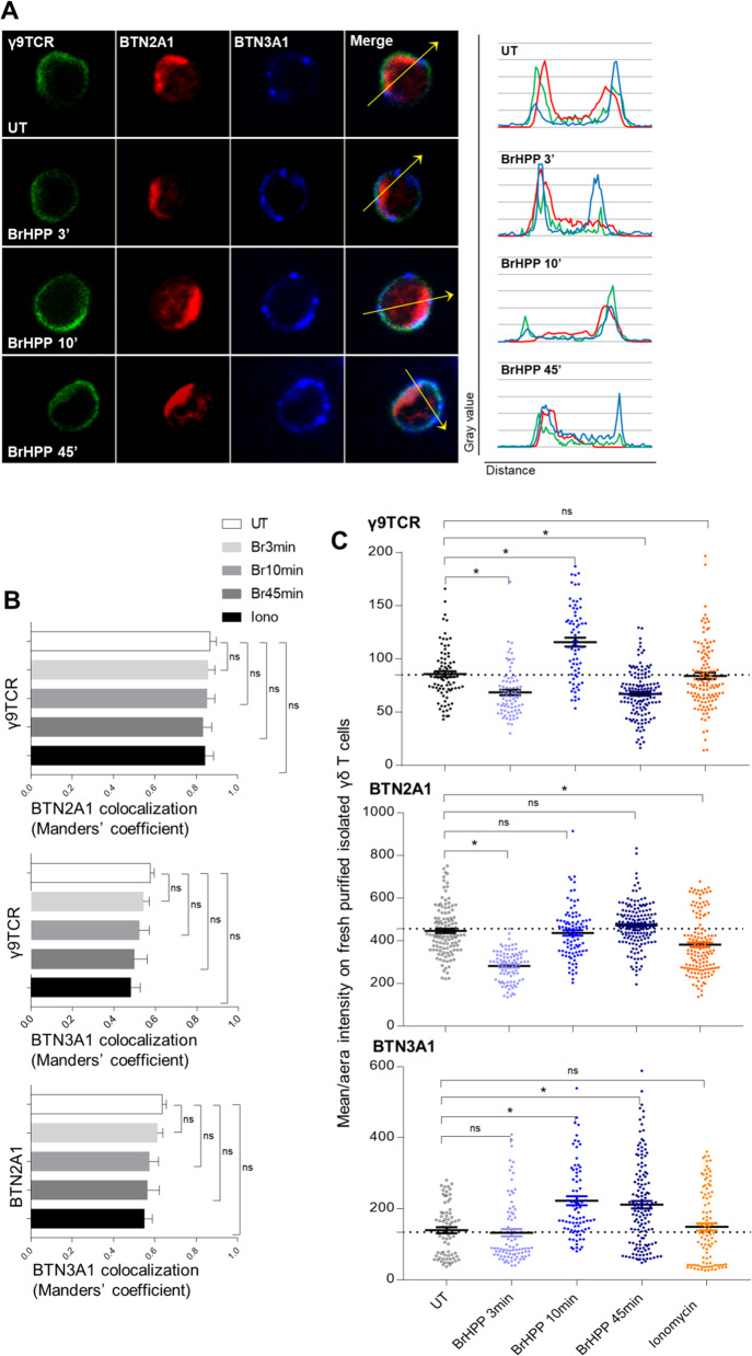
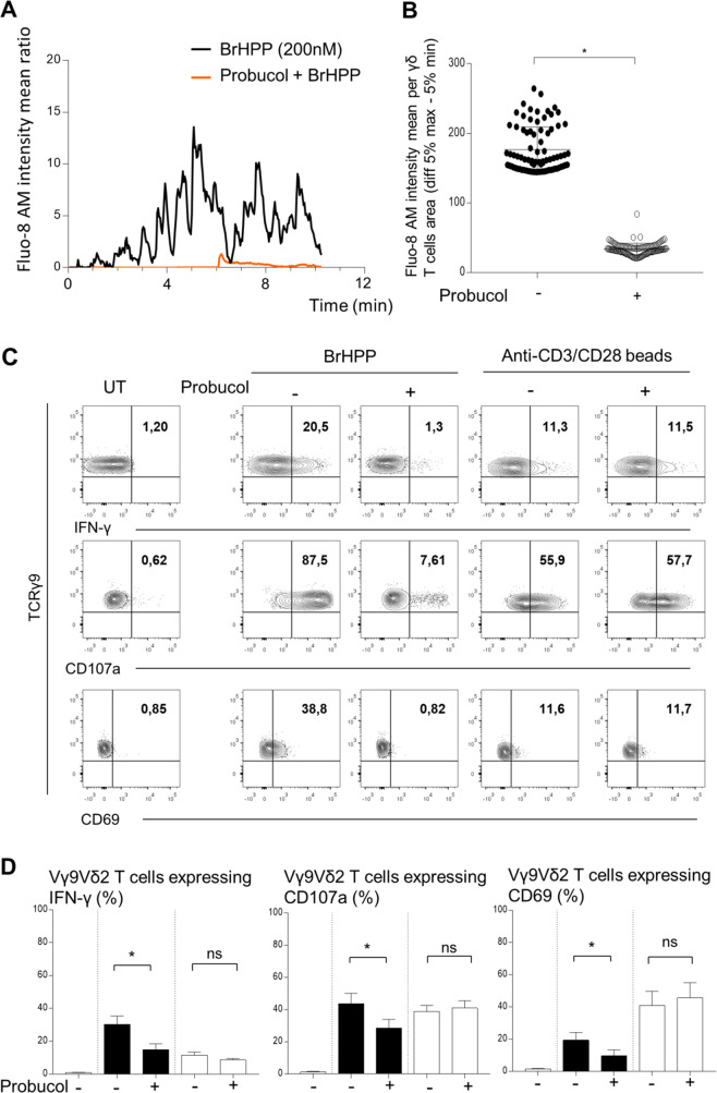
The high cytotoxic activity of Vγ9Vδ2 T lymphocytes against tumor cells makes them useful candidates in anticancer therapies. However, the molecular mechanism of their activation by phosphoantigens (PAgs) is not completely known. Many studies have depicted the mechanism of Vγ9Vδ2 T-cell activation by PAg-sensed accessory cells, such as immune presenting cells or tumor cells. In this study, we demonstrated that pure resting Vγ9Vδ2 T lymphocytes can self-activate through exogenous PAgs, involving their TCR and the butyrophilins BTN3A1 and BTN2A1. This is the first time that these three molecules, concurrently expressed at the plasma membrane of Vγ9Vδ2 T cells, have been shown to be involved together on the same and unique T cell during PAg activation. Moreover, the use of probucol to stimulate the inhibition of this self-activation prompted us to propose that ABCA-1 could be implicated in the transfer of exogenous PAgs inside Vγ9Vδ2 T cells before activating them through membrane clusters formed by γ9TCR, BTN3A1 and BTN2A1. The self-activation of Vγ9Vδ2 T cells, which leads to self-killing, can therefore participate in the failure of γδ T cell-based therapies with exogenous PAgs and should be taken into account.

Keywords: Butyrophilins; Phosphoantigen; T-cell receptor; Vγ9Vδ2 T cells.


Conflict of interest statement

The authors declare no competing interests.

Figures

Fig. 1
Fig. 1
Autologous killing of purified Vγ9Vδ2 T cells requires PAg activation. A Flow chart of the Vγ9Vδ2 T-cell preparation for coculture experiments (γδ R: CMTMR+ γδ T cells; γδ RBr: CMTMR+ γδ T cells pretreated with BrHPP; γδ GBr: PKH67+ γδ T cells pretreated with BrHPP). B, C Flow cytometry analysis of CD107a expression by either γδ RBr or γδ R cocultured with γδ GBr or by γδ GBr cocultured with γδ RBr or γδ R. B One representative experiment; C Twelve independent experiments included CD107a expression by γδR cells cocultured with Daudi cells (D)). D, E Flow cytometry of PKH67 fluorescence acquisition by γδ R or γδ RBr after coculture for 5 min or 4 h with γδ GBr or with PKH67+ Daudi cells (D G). (D One representative experiment (5 min of coculture gave the same result for γδ R or γδ RBr); E seven independent experiments). F, G Flow cytometry analysis of 7-AAD and DAPI staining of γδ RBr or γδ R cells cocultured with γδ GBr cells. (F One representative experiment. G Three independent experiments). Asterisk (*) indicates p < 0.05, Student’s paired t test; ns: not significant
Fig. 2
Fig. 2
Self-activation of resting purified Vγ9Vδ2 T cells by exogenous BrHPP is dependent on TCR, BTN3A1, and BTN2A1. A Sequence of actions for calcium flux detection by video in an individual Vγ9Vδ2 T cell. B–E Time lapse of the Fluo-8 AM intensity representing the calcium flux in one Vγ9Vδ2 T-cell stimulated by ionomycin (B) or exogenous BrHPP (C). Three images were extracted from the time lapses at three different time points of the stimulation. D Mean Fluo-8 AM intensity per γδ T cell area (difference 5% max–5% min). F Time lapse of the Fluo-8 AM intensity representing the calcium flux in one Vγ9Vδ2 T-cell stimulated by BrHPP in the presence or absence of blocking antibodies against γ9TCR, BTN3A1. or BTN2A1. Asterisk (*) indicates p < 0.05, Student’s paired t test; ns not significant
Fig. 3
Fig. 3
Preexisting clusters of γ9TCR, BTN2A1 and BTN3A1 at the surface of resting Vγ9Vδ2 T cells and their modulation during BrHPP stimulation. A Immunofluorescence of γ9TCR, BTN2A1 and BTN3A1 on freshly purified isolated Vγ9Vδ2 T cells during BrHPP stimulation (representative images, γ9TCR: green fluorescence, BTN2A1: red fluorescence and BTN3A1: blue fluorescence and the merge) and colocalization profiles for each condition corresponding to the arrow). B Comparison of the colocalization of γ9TCR, BTN2A1 and BTN3A1 on freshly purified isolated Vγ9Vδ2 T cells during BrHPP or ionomycin stimulation quantified by Manders’ coefficient in ImageJ software. C Mean/area intensity of γ9TCR, BTN2A1, and BTN3A1 on freshly purified isolated Vγ9Vδ2 T cells during BrHPP or ionomycin stimulation. Asterisk (*) indicates p < 0.05, Student’s paired t test; ns not significant
Fig. 4
Fig. 4
Inhibition of ABCA-1 by probucol impairs BrHPP self-activation of resting purified Vγ9Vδ2 T cells. A Time lapse of the Fluo-8 AM intensity representing the calcium flux in one Vγ9Vδ2 T-cell stimulated by BrHPP and previously treated with Probucol (10 µM, overnight). B Mean Fluo-8 AM intensity per γδ T-cell area (difference 5% max–5% min). C, D Flow cytometry analysis of the expression of CD69 and CD107a and IFN-γ production by fresh purified Vγ9Vδ2 T cells activated by BrHPP or anti-CD3/CD28 beads previously treated with Probucol (10 µM, overnight). (C A representative experiment; D Four independent experiments; black: Vγ9Vδ2 T cells activated by BrHPP; white: Vγ9Vδ2 T cells activated by anti-CD3/CD28 beads). Asterisk (*) indicates p < 0.05, Student’s paired t test; ns not significant

References

    1. Choudhary A, Davodeau F, Moreau A, Peyrat MA, Bonneville M, Jotereau F. Selective lysis of autologous tumor cells by recurrent gamma delta tumor-infiltrating lymphocytes from renal carcinoma. J Immunol. 1995;154:3932–40. - PubMed
    1. Kabelitz D, Wesch D, Pitters E, Zöller M. Characterization of tumor reactivity of human V gamma 9V delta 2 gamma delta T cells in vitro and in SCID mice in vivo. J Immunol. 2004;173:6767–76. doi: 10.4049/jimmunol.173.11.6767. - DOI - PubMed
    1. Sicard H, Ingoure S, Luciani B, Serraz C, Fournié J-J, Bonneville M, et al. In vivo immunomanipulation of V gamma 9V delta 2 T cells with a synthetic phosphoantigen in a preclinical nonhuman primate model. J Immunol. 2005;175:5471–80. doi: 10.4049/jimmunol.175.8.5471. - DOI - PubMed
    1. Rei M, Pennington DJ, Silva-Santos B. The emerging Protumor role of γδ T lymphocytes: implications for cancer immunotherapy. Cancer Res. 2015;75:798–802. doi: 10.1158/0008-5472.CAN-14-3228. - DOI - PubMed
    1. Donia M, Ellebaek E, Andersen MH, Straten PT, Svane IM. Analysis of Vδ1 T cells in clinical grade melanoma-infiltrating lymphocytes. OncoImmunology. 2012;1:1297–304. doi: 10.4161/onci.21659. - DOI - PMC - PubMed

Publication types