. 2021 Dec;35(3):853-861.
doi: 10.1007/s12028-021-01233-0. Epub 2021 Jun 28.

Electrocerebral Signature of Cardiac Death

Affiliations

Electrocerebral Signature of Cardiac Death

Adu L Matory et al. Neurocrit Care. 2021 Dec.

Abstract

Background: Electroencephalography (EEG) findings following cardiovascular collapse in death are uncertain. We aimed to characterize EEG changes immediately preceding and following cardiac death.

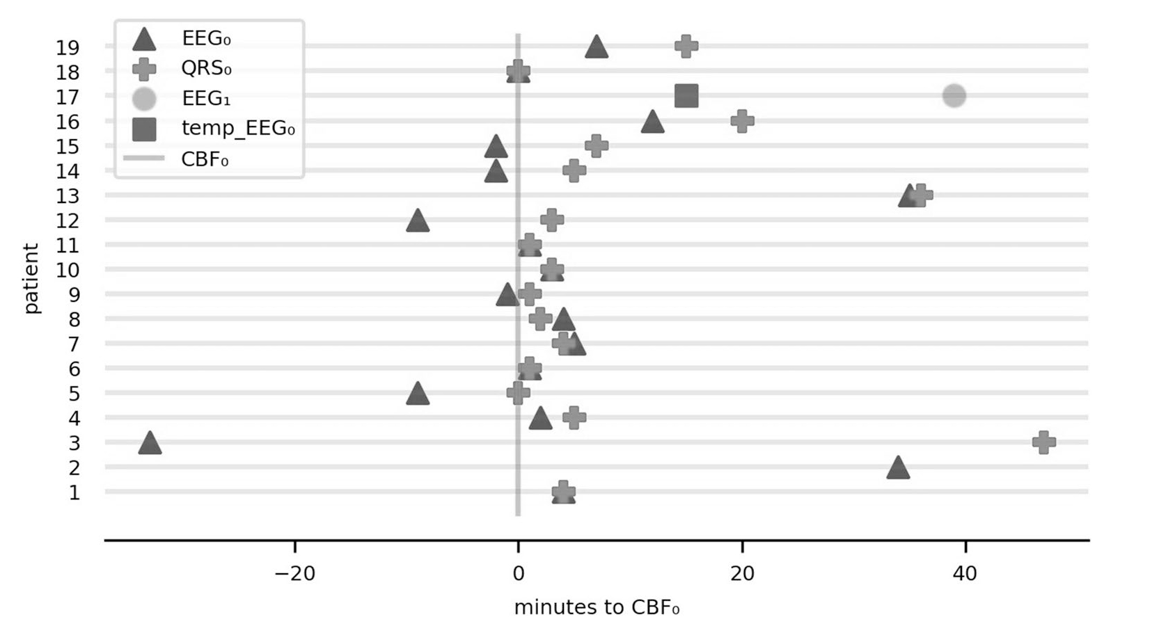
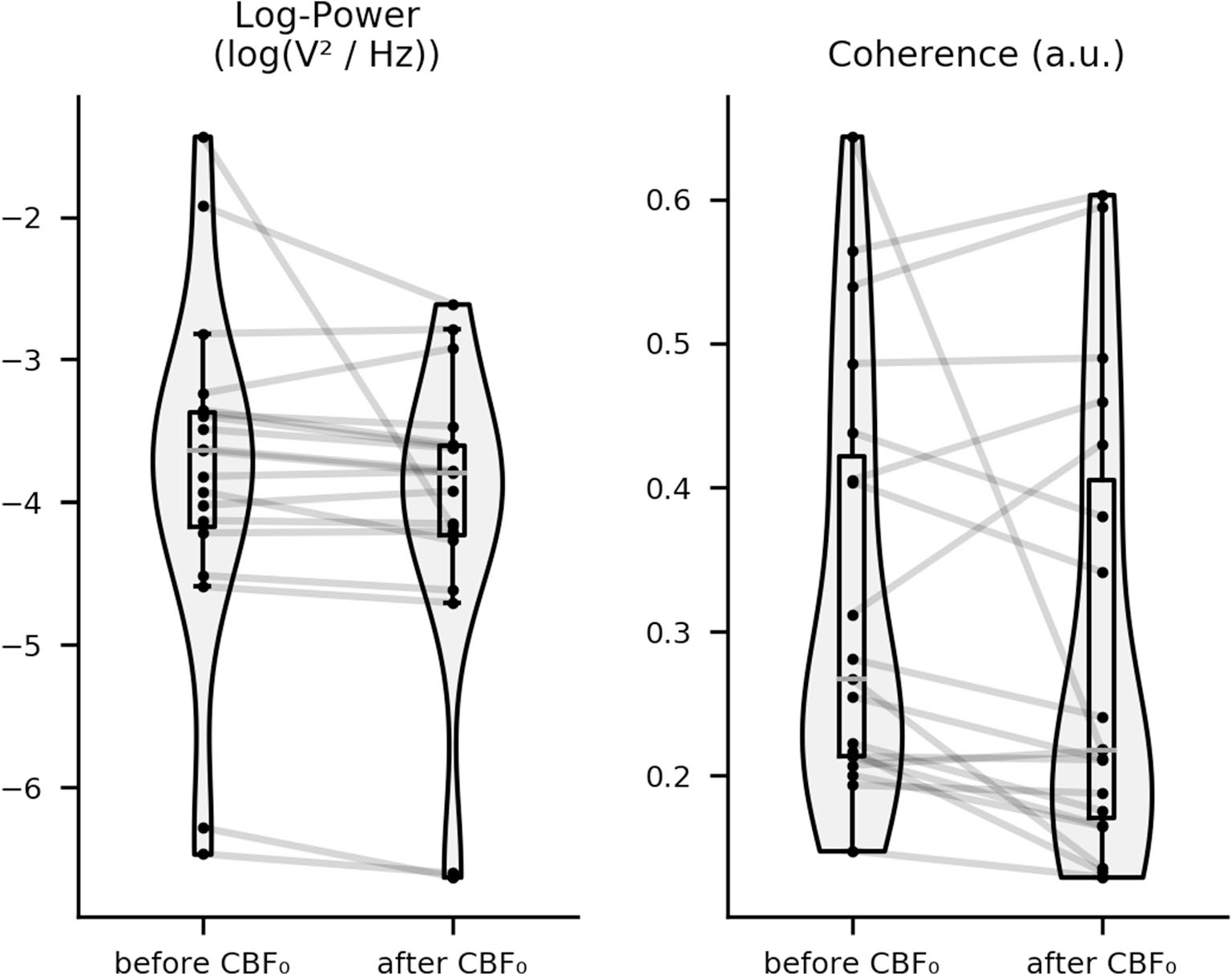
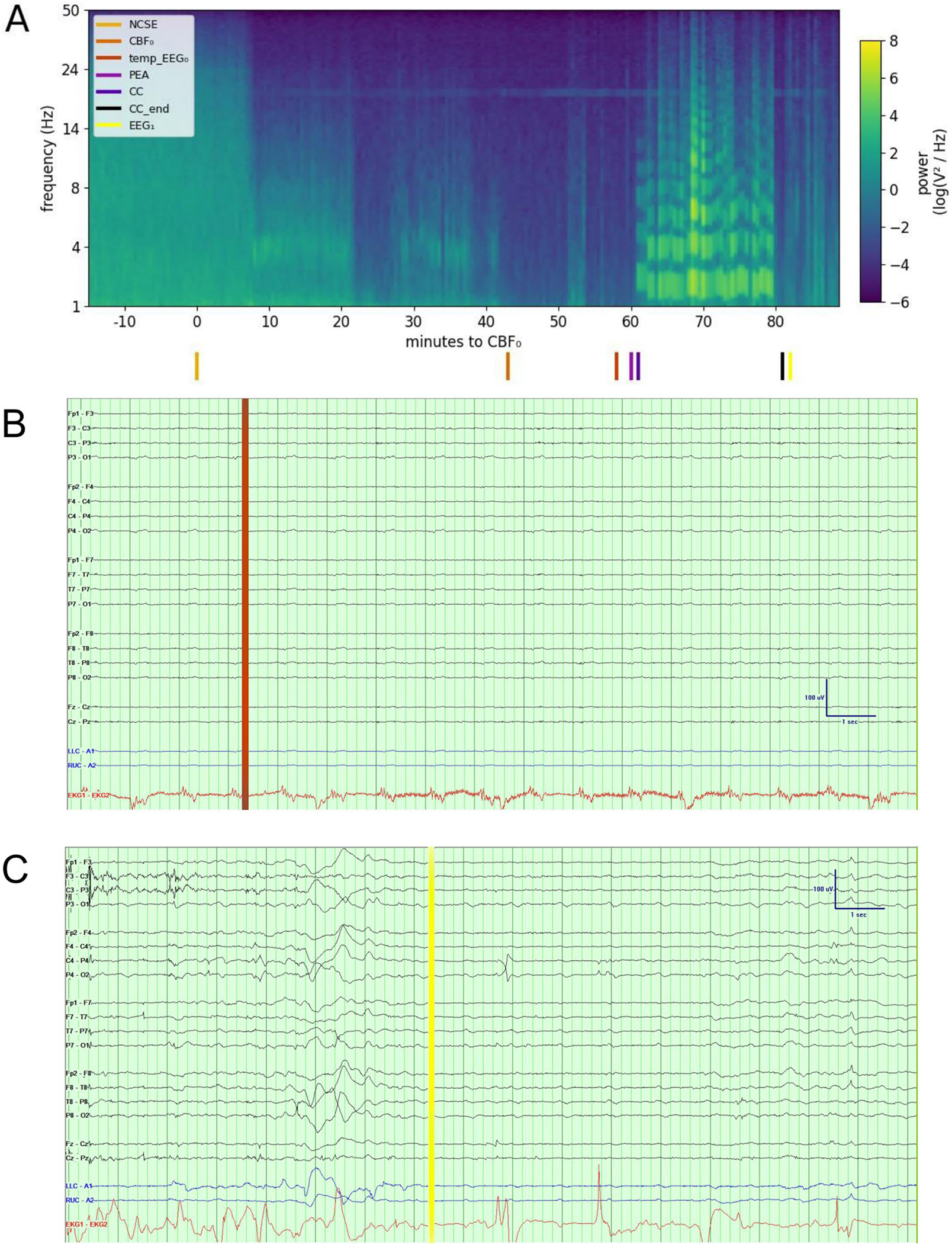
Methods: We retrospectively analyzed EEGs of patients who died from cardiac arrest while undergoing standard EEG monitoring in an intensive care unit. Patients with brain death preceding cardiac death were excluded. Three events during fatal cardiovascular failure were investigated: (1) last recorded QRS complex on electrocardiogram (QRS0), (2) cessation of cerebral blood flow (CBF0) estimated as the time that blood pressure and heart rate dropped below set thresholds, and (3) electrocerebral silence on EEG (EEG0). We evaluated EEG spectral power, coherence, and permutation entropy at these time points.

Results: Among 19 patients who died while undergoing EEG monitoring, seven (37%) had a comfort-measures-only status and 18 (95%) had a do-not-resuscitate status in place at the time of death. EEG0 occurred at the time of QRS0 in five patients and after QRS0 in two patients (cohort median - 2.0, interquartile range - 8.0 to 0.0), whereas EEG0 was seen at the time of CBF0 in six patients and following CBF0 in 11 patients (cohort median 2.0 min, interquartile range - 1.5 to 6.0). After CBF0, full-spectrum log power (p < 0.001) and coherence (p < 0.001) decreased on EEG, whereas delta (p = 0.007) and theta (p < 0.001) permutation entropy increased.

Conclusions: Rarely may patients have transient electrocerebral activity following the last recorded QRS (less than 5 min) and estimated cessation of cerebral blood flow. These results may have implications for discussions around cardiopulmonary resuscitation and organ donation.

Keywords: Brain hypoxia; Cardiac arrest; Consciousness; Death; Encephalography; Hypotension.


Conflict of interest statement

Conflicts of Interest

JC reports grants from the National Institute of Neurological Disorders and Stroke and the Dana Foundation. He is a minority shareholder at iCE Neuro-systems. None of these constitute a conflict of interest to the work presented here. The remaining authors do not have any conflicts of interest.

Figures

Fig. 1
Fig. 1
Timeline of three pathophysiologic events. Relationship of electrocerebral silence on EEG (EEG0) and last QRS complex on ECG (QRS0) in relation to cessation of cerebral blood flow (CBF0). The y axis represents the 19 patients in the study and the x axis represents time in minutes. Patients are ordered by magnitude of change in global power 5 min before vs. 5 min after CBF0. For patient 12, temp_EEG0 represents time of temporary electrocerebral silence before the return of cerebral activity (EEG1; see Fig. 3 for details). QRS0 was not available in two patients (no. 2 and no. 17) because of ECG artifacts following chest compressions and poorly connected ECG leads. ECG electrocardiogram, EEG electroencephalograph, EEG1 return of cerebral activity
Fig. 2
Fig. 2
Change in full-spectrum EEG features after CBF0. Boxplot showing change in epoch-averaged and channel-averaged full-spectrum EEG features 5 min before vs. 5 min after CBF0 for all patients. Cohort-wide changes in log power and coherence are statistically significant (p < 0.028). CBF0 cessation of cerebral blood flow, EEG electroencephalograph
Fig. 3
Fig. 3
Raw EEG data and power spectral density in a patient with electrocerebral activity on EEG after CBF0 and a brief period of isoelectric EEG. Sixty-five-year-old woman who was hospitalized for refractory status epilepticus in the setting of anoxic brain injury. On hospital day 10, the course was complicated by cardiac arrest with PEA. After chest compressions were performed for 20 min, the patient underwent withdrawal of life-sustaining therapies at the request of the family. Power spectral density of her EEG is displayed (a), calculated from channels Fz, Cz, and Pz because these are less frequently affected by muscle and ECG artifact. Colored lines at the bottom indicate nonconvulsive status epilepticus (NCSE), CBF0, EEG0, PEA, start of chest compressions (CC), end of chest compressions (CC_end), and return of cerebral activity ( EEG1). EEG (bipolar montage, sensitivity of 5 μV/mm, 60-Hz notch filter) at the time (temp_EEG0) demonstrated no clear electrocerebral activity (b). Following the activity after CBF0 and a brief period of isoelectric EEG, electrocerebral activity returned ( EEG1) (c) (see Supplemental Fig. 2 for raw EEG data at the remaining events). The power spectral density plot revealed decreased power 36 min before CBF0. Artifacts are seen throughout in the 20-Hz range, and prominent artifacts due to chest compressions are seen between 18 and 38 min following CBF0. At CBF0, median log power was − 4.10 (IQR − 12.08 to − 2.69, range − 16.61 to 2.96), median coherence was 0.23 (IQR 0.01–0.94, range − 0.50 to 0.99), and median permutation entropy was 3.31 (IQR 2.60–3.87, range 1.85–4.45). CBF0 cessation of cerebral blood flow, ECG electrocardiogram, EEG electroencephalograph, EEG0 electrocerebral silence on EEG, IQR interquartile range, PEA pulseless electrical activity

References

    1. Salehi S, Tran K, Grayson WL. Advances in perfusion systems for solid organ preservation. Yale J Biol Med. 2018;91(3):301–12. - PMC - PubMed
    1. Blundon EG, Gallagher RE, Ward LM. Electrophysiological evidence of preserved hearing at the end of life. Sci Rep. 2020;10(1):10336. - PMC - PubMed
    1. Pozhitkov AE, Neme R, Domazet-Lošo T, et al. Tracing the dynamics of gene transcripts after organismal death. Open Biol. 2017;7(1):160267. - PMC - PubMed
    1. Ferreira PG, Muñoz-Aguirre M, Reverter F, et al. The effects of death and post-mortem cold ischemia on human tissue transcriptomes. Nat Commun. 2018;9(1):490. - PMC - PubMed
    1. Borjigin J, Lee U, Liu T, et al. Surge of neurophysiological coherence and connectivity in the dying brain. Proc Natl Acad Sci. 2013;110(35):14432–7. - PMC - PubMed

Publication types